

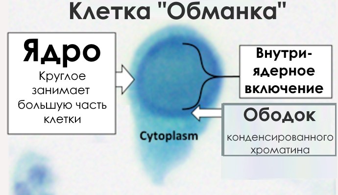
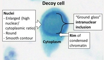
🔬 Urine Sediment Cytology ✅ A high nuclear-to-cytoplasmic ratio makes it resemble a cancer cell (hence the name “decoy cell”), but the presence of an inclusion body distinguishes it.
❓ Decoy cells in cytology: what is important to know?
Decoy cells are virus-infected urothelial cells predominantly found in urine. They are more common in immunocompromised patients, such as transplant recipients, HIV patients, or other immunodeficient groups. Their name is associated with their characteristic morphology, which resembles cancer cells, making diagnosis difficult.
Cytologically, decoy cells are characterized by an enlarged nucleus with a basophilic intranuclear inclusion surrounded by chromatin. This chromatin gives the nucleus a “ground-glass” or “jelly-like” appearance. Sometimes, inclusions may be vesicular with a characteristic halo. These features can be identified with Papanicolaou staining, as well as using phase-contrast microscopy.
Decoy cells are most often found in urine sediment during cytological examination. In rare cases, they can also be detected in kidney biopsy samples, indicating polyomavirus-associated nephropathy (BKVAN), or in respiratory secretions or other biological fluids during active viral infection.
These cells have clinical significance, as their presence may indicate viral activity. In kidney transplant recipients, they are associated with BK polyomavirus nephropathy, and in hematopoietic stem cell transplant recipients, with hemorrhagic cystitis. However, decoy cells themselves do not cause pathology.
Treatment is aimed at eliminating viral activity and includes reducing the dose of immunosuppressants in transplant recipients or using antiretroviral therapy in HIV patients. For cytologists, decoy cells serve as an important diagnostic marker, allowing differentiation of viral infection from oncological diseases and assessment of the condition of immunocompromised patients.


