

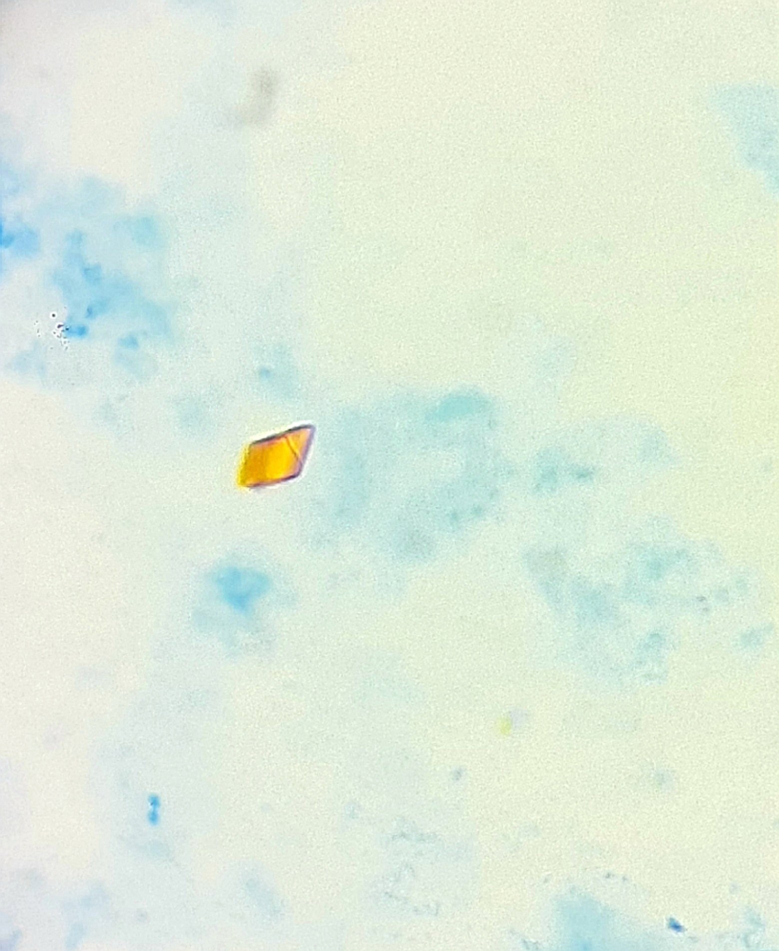
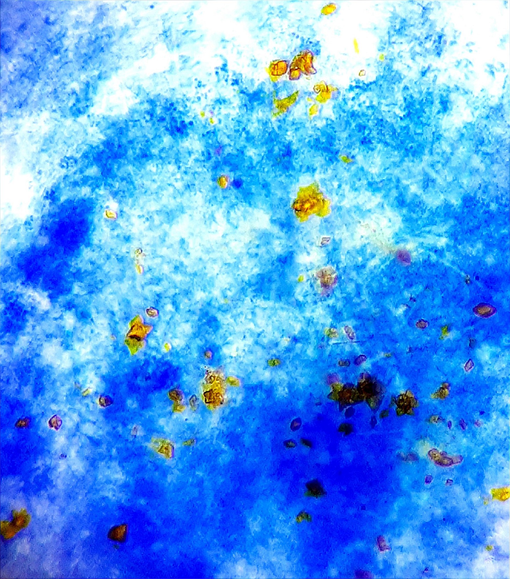
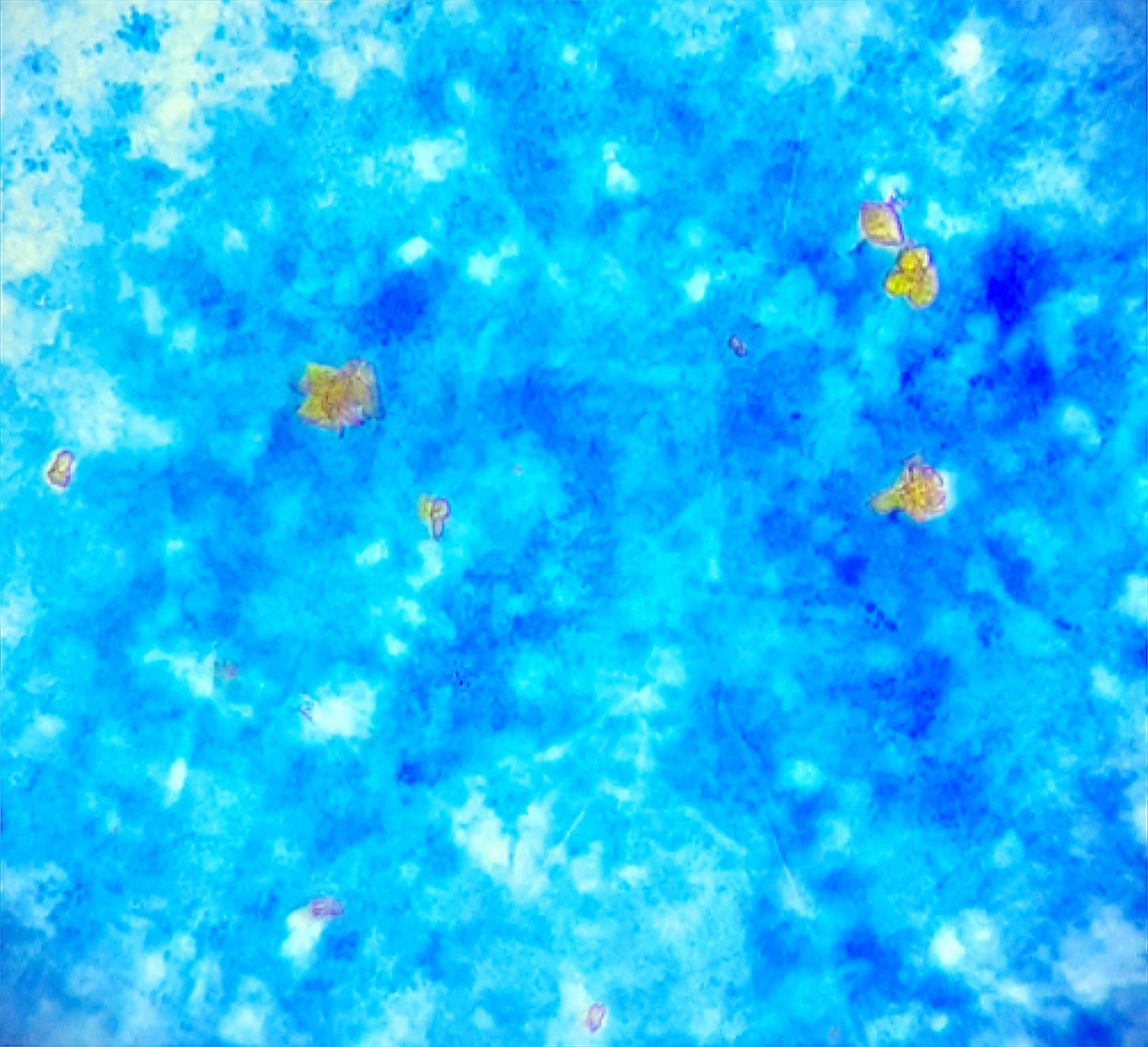
Hematoidin Crystals in Pleural Fluid 🔬
🙏 The clinical case was kindly provided by Vera Kirnas, Biologist, Clinical Diagnostic Laboratory, F.S. Ankudinov Yuzhno-Sakhalinsk City Hospital.
👉 Hematoidin crystals in pleural fluid indicate the long-term presence of blood in the cavity. Hematoidin is a pigment formed from hemoglobin under low oxygen conditions and the absence of active blood cells. The main causes of hematoidin crystals in pleural fluid may include:
✅ Chest or organ trauma, leading to hemorrhage into the pleural cavity. ✅ Malignant processes (e.g., pleural metastases in lung cancer, breast cancer, or other tumors) that can cause hemorrhage into the pleural cavity. ✅ Hemothorax, especially when detected late or with prolonged blood retention in the pleural cavity without timely drainage. ✅ Pulmonary infarction or other ischemic processes that lead to tissue destruction and hemorrhage. ✅ Pleural tuberculosis, especially in the chronic stage, where tissue destruction and prolonged inflammation with hemorrhage occur.
Thus, the detection of hematoidin often indicates that blood has been present in the pleural cavity for a long time.


