

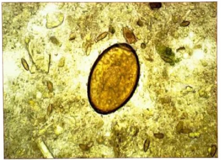
👉The nanophyetus egg is golden in color, with a clearly visible operculum and a wide, slightly protruding knob above the eggshell. The eggshell is rough. Magnification x630
✅The disease is caused by the small trematode Nanophyetus schikhobalowi.
✅Mature individuals parasitize the small intestine. In the natural environment, definitive hosts are fish-eating predatory animals. The development of this biohelminth occurs with a change of two intermediate hosts – a mollusk and freshwater fish of the salmon family.
✅Natural foci of nanophyetiasis in Russia are located in the Amur and Ussuri river basins, and on Sakhalin. Human infection occurs by consuming thermally unprocessed fish infected with metacercariae.
✅The leading symptoms of the invasion are abdominal pain and unstable stools with a tendency to diarrhea. In cases of abundant invasion, severe and intractable ulcerative enteritis may develop. Leukocytosis and peripheral blood eosinophilia are not observed in sparse invasions and uncomplicated courses. The presence of an epidemiological history is important in the diagnosis of nanophyetiasis.
✅Nanophyetus eggs, found in fecal matter using enrichment methods, resemble Diphyllobothrium latum eggs. For confident differentiation of Nanophyetus and Diphyllobothrium latum eggs, it is necessary to examine them under high magnification.
Source: Mironova L.A. - General Clinical Studies, 2021


