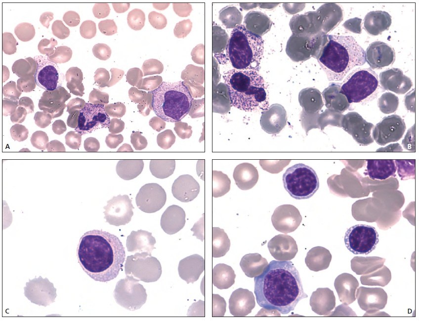
Large granular lymphocyte leukemia. Peripheral blood smear. (A) On the left, a lymphoid cell with abundant pale cytoplasm containing numerous small granules; the nucleus is compact, with chromatin condensed into blocks. On the right, a large granular lymphocyte (LGL) that appears less mature, with more abundant pink cytoplasm; the nucleus is large, indented and curved with a small degree of chromatin condensation. (B) On the left, a metamyelocyte and a myelocyte. On the right, two large granular lymphocytes with varying amounts of cytoplasm. (C) This cell, in which granules are not identified, is also an LGL, as indicated by the abundant pale lilac-colored cytoplasm. (D) This cell population demonstrates some heterogeneity: at the bottom, a large cell with abundant basophilic cytoplasm, the cell is quite fragile as it has been deformed by surrounding red blood cells. The other two cells have no visible granules: on the right there is a ring of small vacuoles. The chromatin structure, however, is similar to that of other LGLs shown in (A - D). Source: Giuseppe d’Onofrio - Morphology of Blood Disorders 2015
Large Granular Lymphocyte Leukemia in Blood Smear
Morphological features of large granular lymphocytes in LGL leukemia blood smear: nuclear atypia, cytoplasmic granularity, and cellular heterogeneity.