

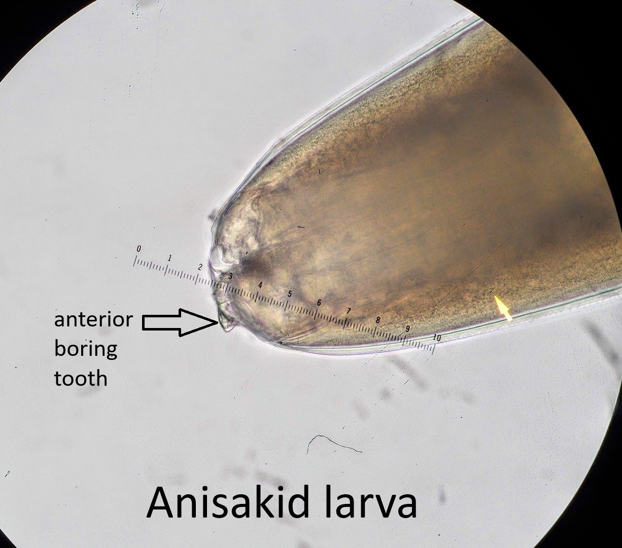
Answer to the parasitology challenge: Anisakid larva
The size and characteristic morphological features of this worm — including the anterior perforating (“boring”) tooth and a slightly rounded caudal end with a small mucron — along with the “fish” history, indicate that this is a third-stage (L3) anisakid larva.
Several readers wished the parasitologist who found the anisakid “bon appétit,” to which he replied that he “preserved the worm in formalin for study, and caramelized the worm-free fillet with chopped green onions, ground black pepper, garlic, cayenne pepper, and fish sauce in a clay pot for dinner with rice. It was very delicious, and he felt no gastrointestinal discomfort.” This is dedication to parasitology — and an excellent reminder that finding one worm does not necessarily mean that all the other fish should be discarded!
Anisakiasis is a zoonotic infection caused by third-stage (L3) ascaridoid nematode larvae, most commonly species of the genera Anisakis, Pseudoterranova, and Contracaecum. Humans become accidental hosts by consuming raw or undercooked fish or squid containing viable larvae. Since humans are not suitable definitive hosts, the larvae cannot reach the adult stage, but they can penetrate the gastric or intestinal mucosa and cause disease.
Anisakid larvae recovered from clinical specimens are typically several centimeters long (~20–35 mm) and can be presumptively identified by macroscopic and microscopic morphology. At the anterior end, there is a small triangular boring tooth, which the larva uses to penetrate host tissues. Note the developing lips; adult ascaridoid nematodes have 3 well-developed fleshy lips. Near the anterior end, there is also an excretory pore, the location of which can be useful for identification. The boring tooth can be difficult to discern and may be damaged or lost during worm extraction from the mucosa during upper endoscopy.
The posterior end provides another useful clue. Some Anisakid L3 larvae have a small terminal projection called a mucron, which is clearly visible in a well-preserved specimen, as in this case. The presence or absence of the mucron, along with features of the digestive tract and the location of the excretory pore, helps distinguish the main anisakid groups. The mucron is typically present in Pseudoterranova and Anisakis Type I larvae, whereas it is absent in Anisakis Type II and Contracaecum.
However, species identification based solely on larval morphology can be challenging, and molecular methods are usually required for definitive identification.


