
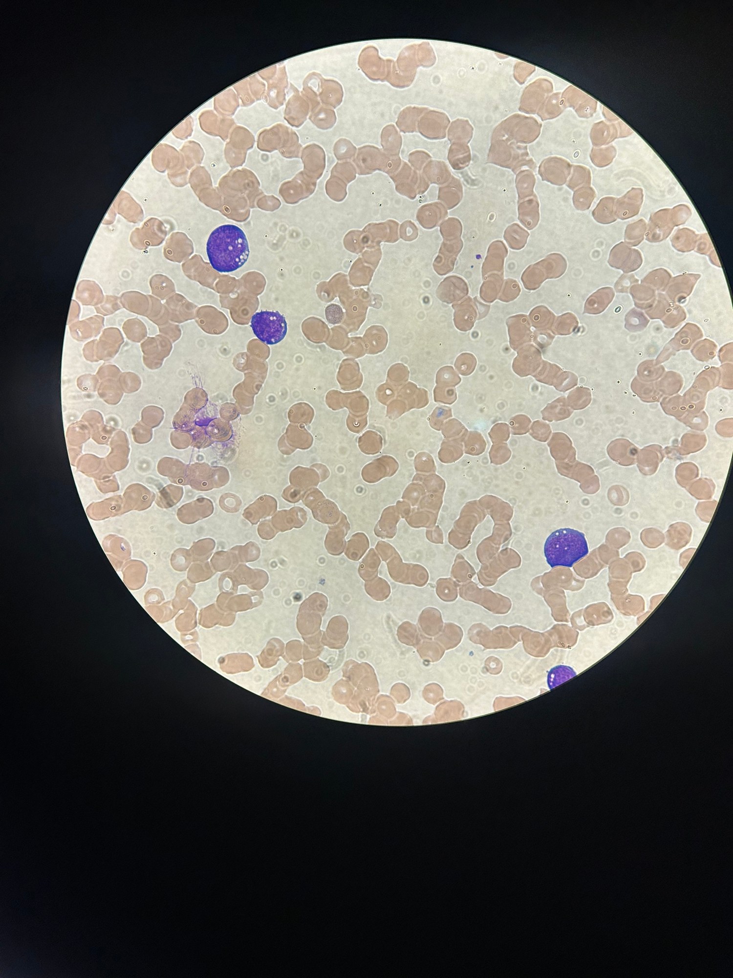
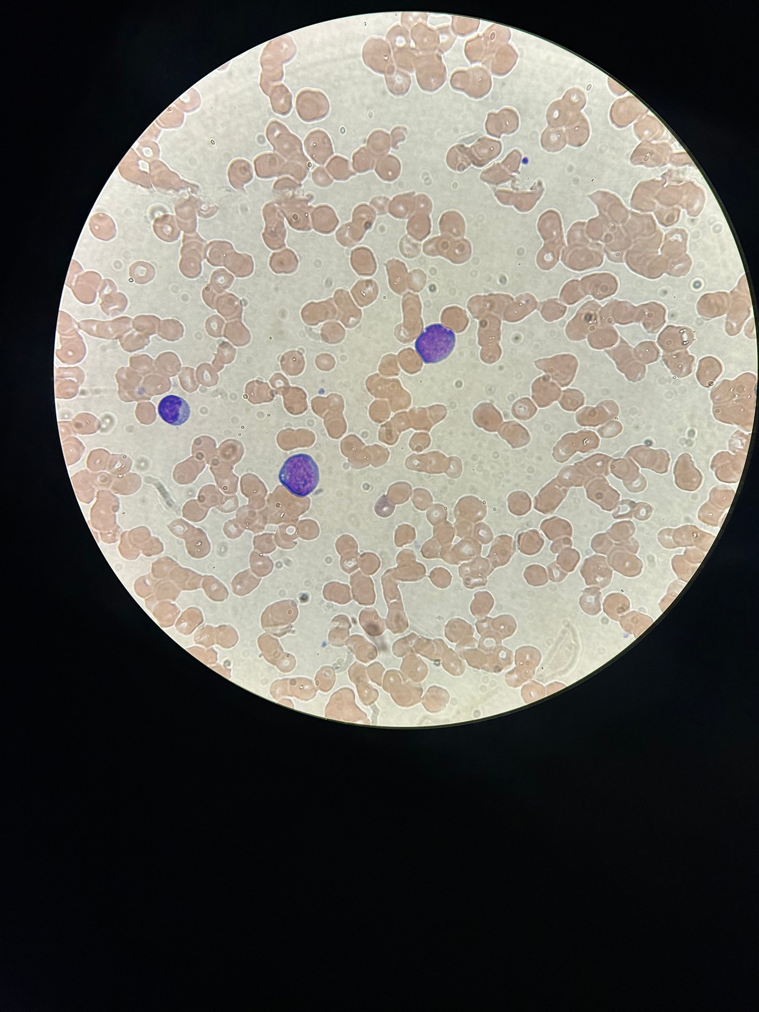
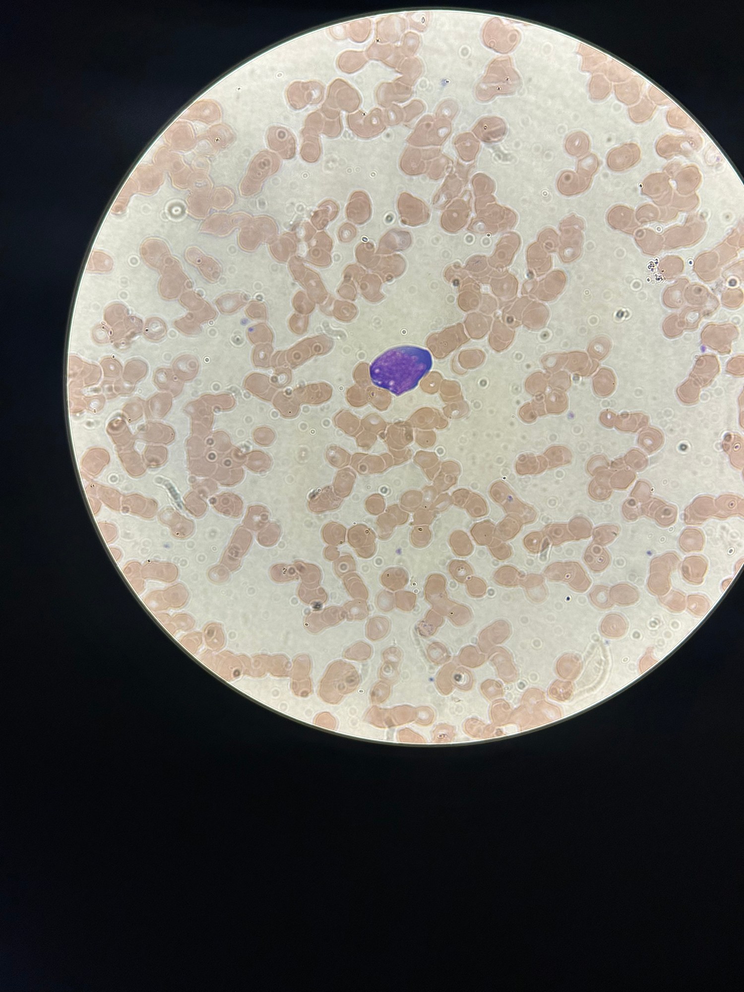
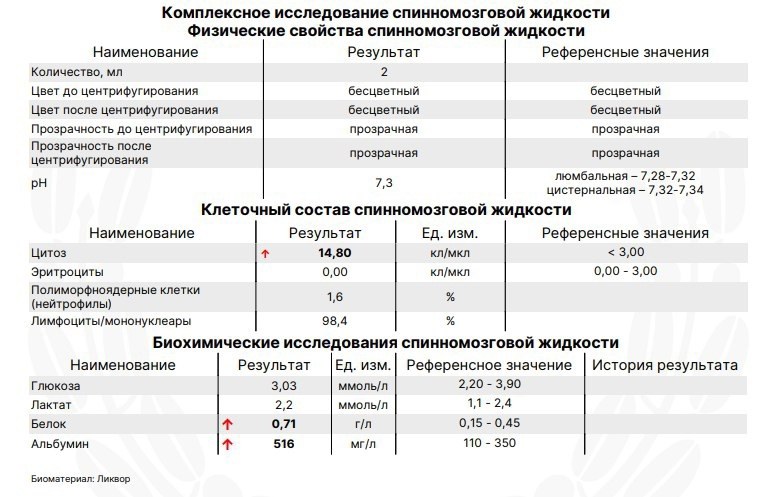
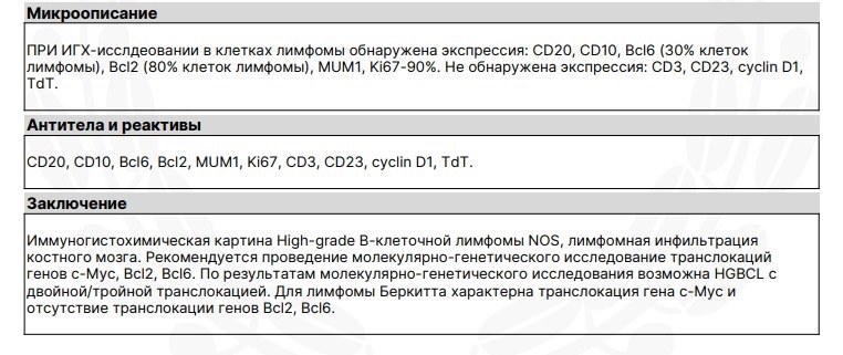
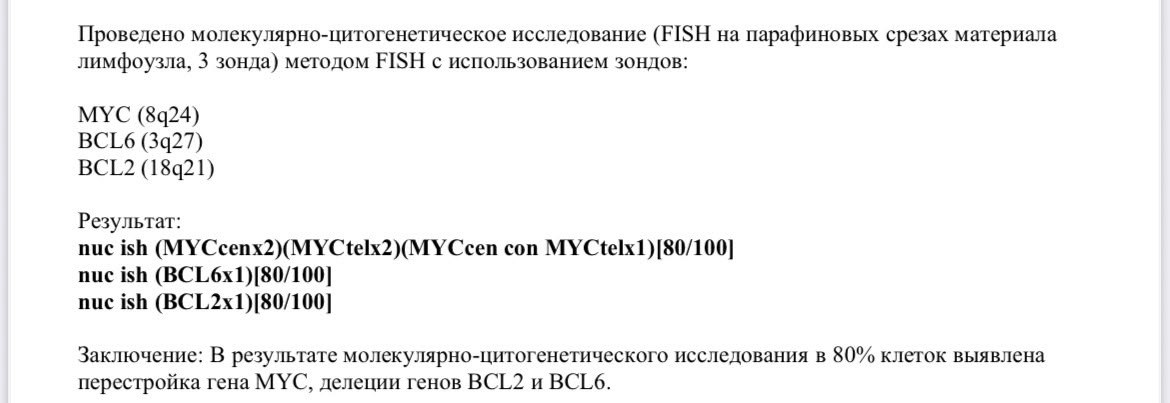
Clinical case: Burkitt lymphoma with central nervous system (CNS) involvement.
Cell morphology (cytological characteristics): ✅ Nuclei: round, sometimes with cleavage (indentation). ✅ Chromatin: finely granular (delicately dispersed). ✅ Nucleoli: may be present, usually few. ✅ Cytoplasm: intensely basophilic, forming a narrow rim around the nucleus. A characteristic but inconstant feature is vacuolization, due to inclusions of neutral lipids.
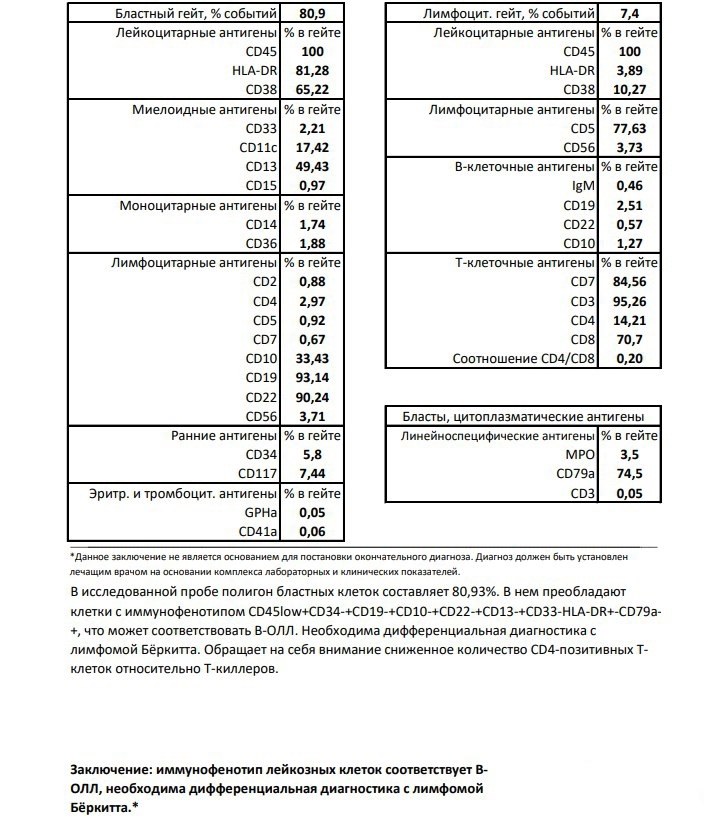
💡 The immunophenotype of Burkitt lymphoma is similar to that of mature B-cell acute lymphoblastic leukemia (B-ALL) (L3 stage according to FAB classification).
Differential diagnosis: ✅ Accurate diagnosis verification requires additional studies: 🔬 Cytogenetic analysis (FISH) and 🧬 Molecular genetic analysis The main goal: detection of translocation involving the c-MYC gene (most often t(8;14)), which is a key diagnostic criterion and allows exclusion of other mature B-cell lymphomas and leukemias.
👉 Material kindly provided by clinical laboratory diagnostics physician Alena Leshchiner, Hadassah Medical Moscow.


