

🔬 Lymphocyte-predominant Hodgkin lymphoma, imprint smears from a cervical lymph node, Romanowsky-Giemsa stain
🙏 Smear photographs kindly provided by Kristina Yuryevna, Clinical Laboratory Diagnostics Physician, BU “RCB”, Cheboksary
Cytological features of lymphocyte-predominant Hodgkin lymphoma: microscopy of imprint smears from a cervical lymph node, Romanowsky-Giemsa stain.
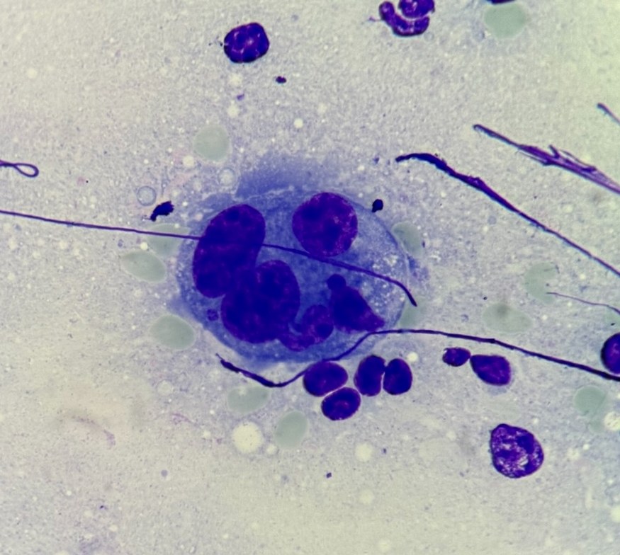
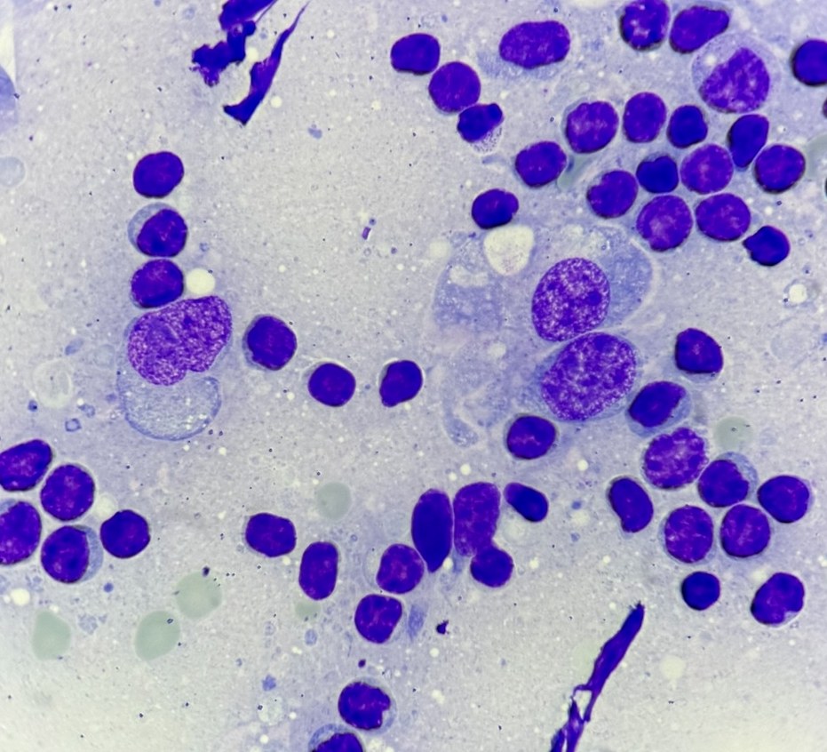
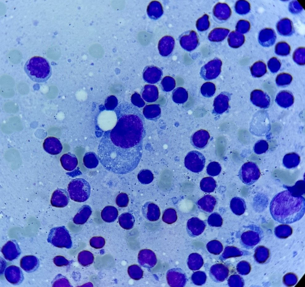
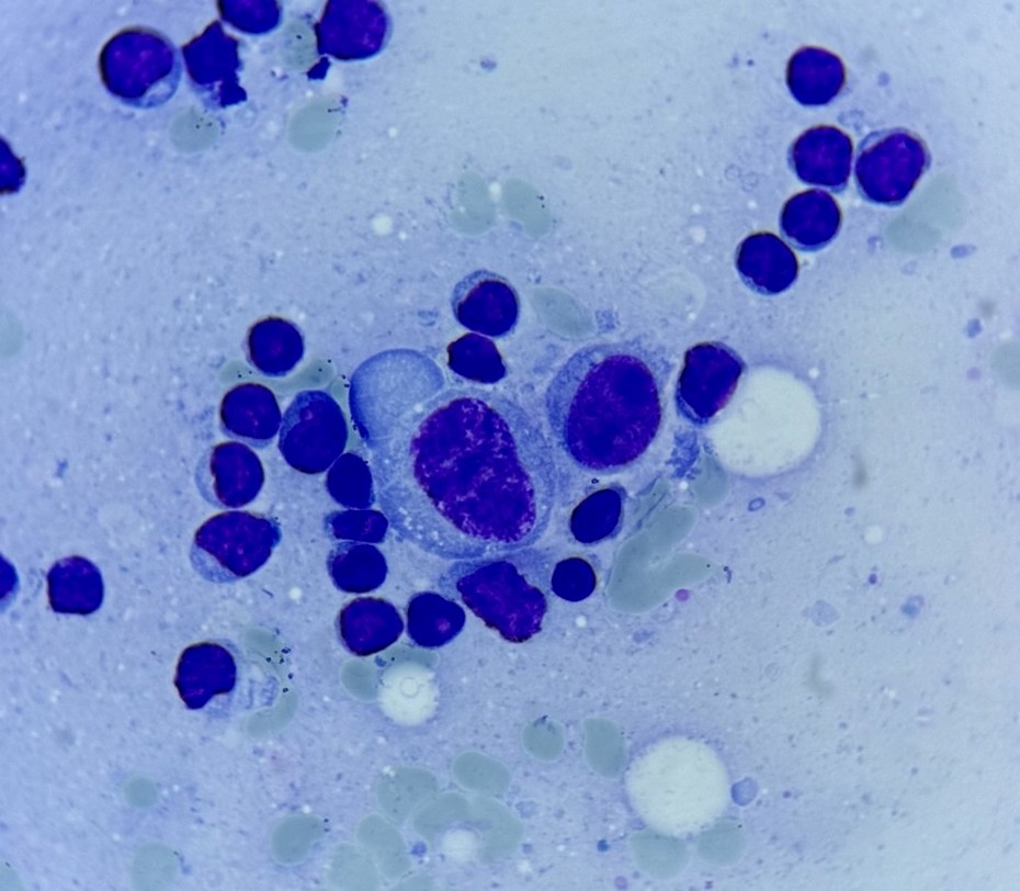
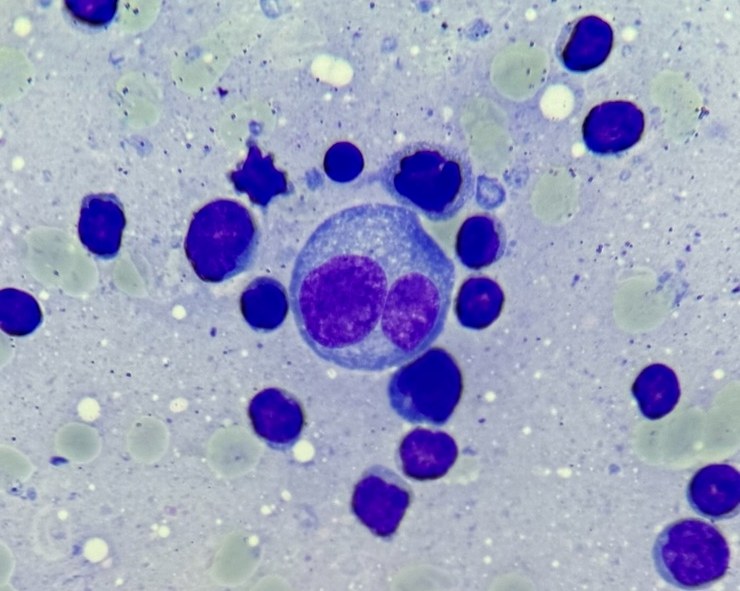
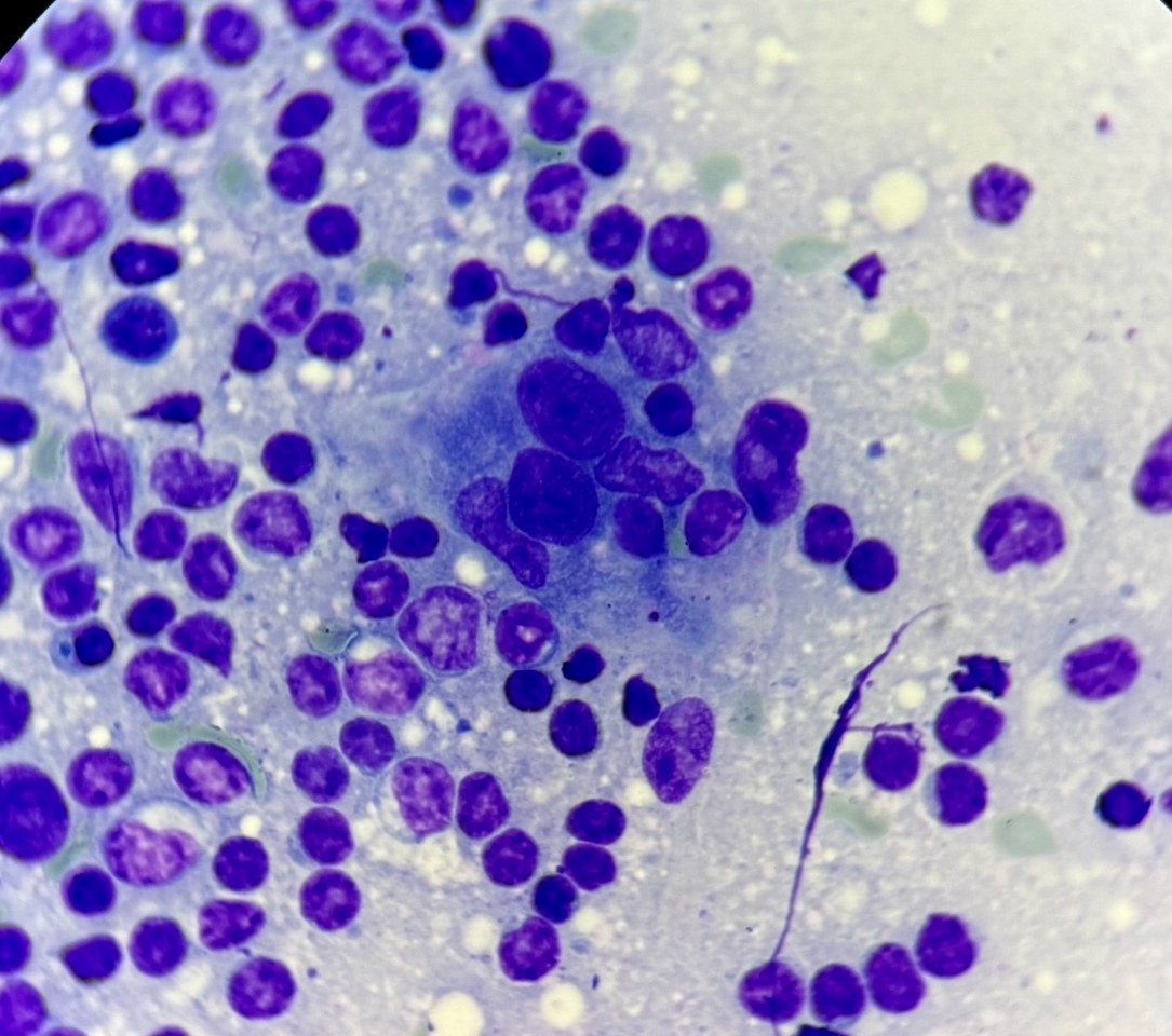
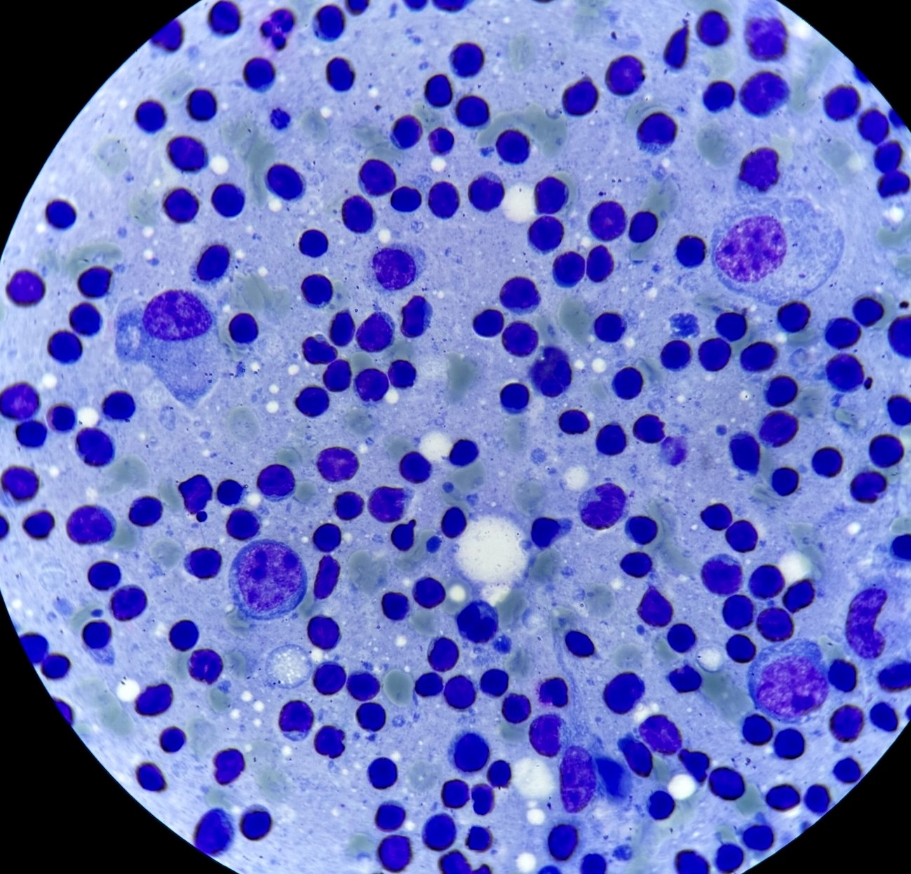
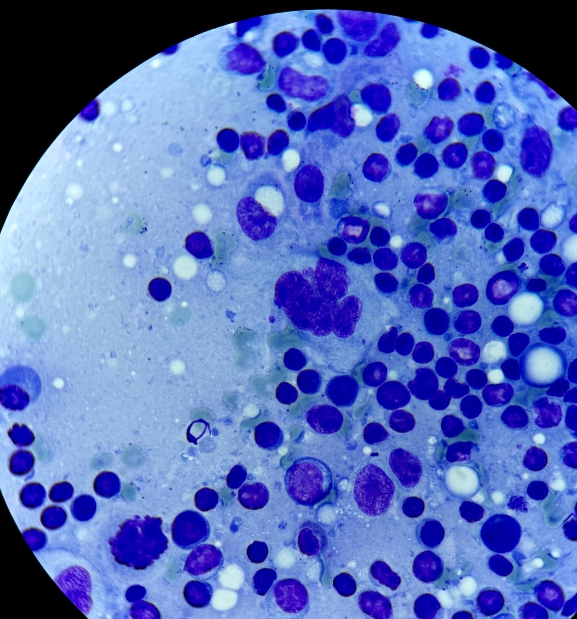
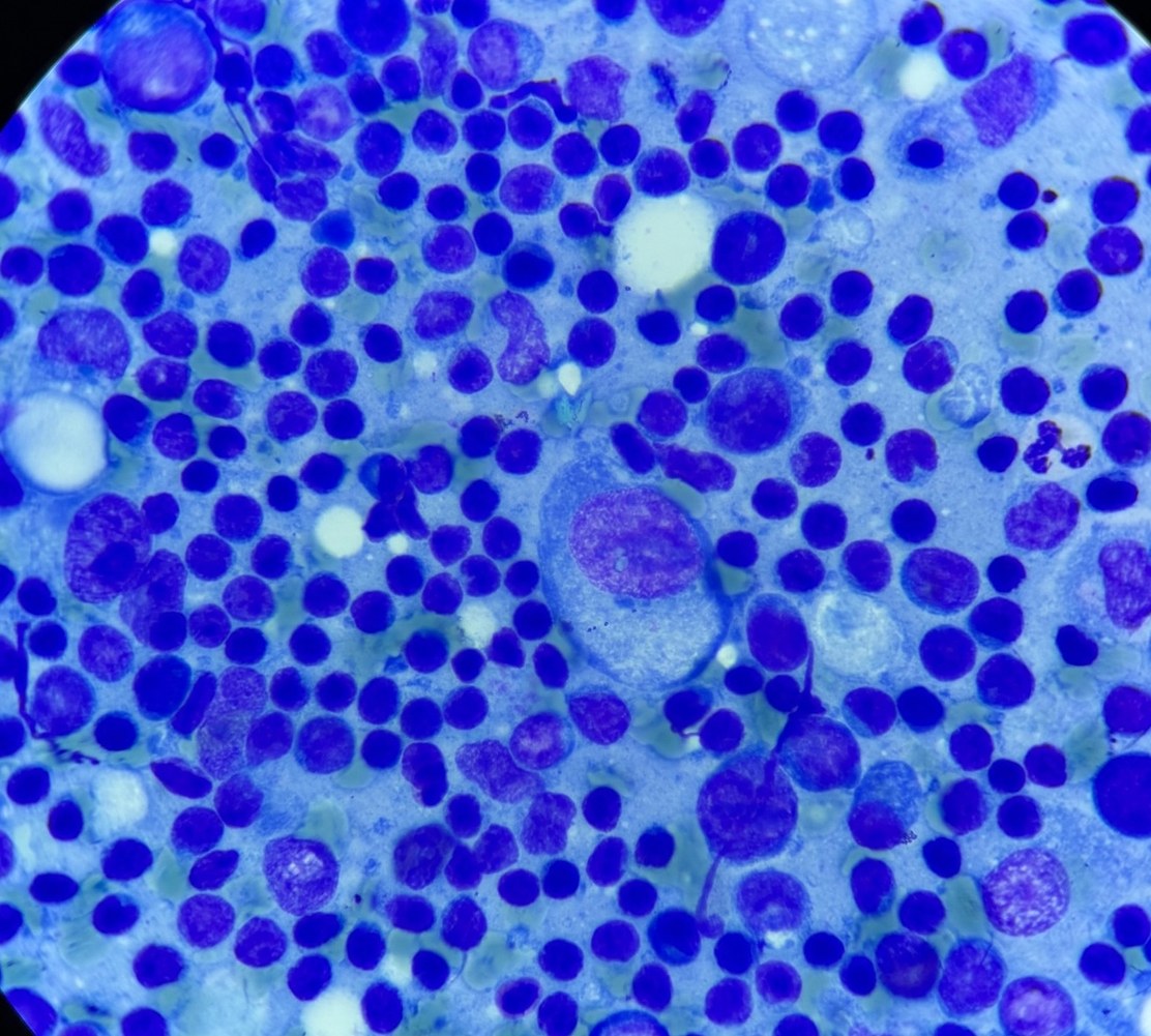
🔬 Lymphocyte-predominant Hodgkin lymphoma, imprint smears from a cervical lymph node, Romanowsky-Giemsa stain
🙏 Smear photographs kindly provided by Kristina Yuryevna, Clinical Laboratory Diagnostics Physician, BU “RCB”, Cheboksary

Cytological features of a lymph node impression smear in Hodgkin lymphoma: visualization of a blood capillary, endothelial cells, and erythrocytes.

A clinical case of Hodgkin lymphoma: nodular sclerosis. Cytology of a biopsy touch imprint: Hodgkin and Reed-Sternberg cells among lymphocytes.

Cytological features of a lymph node imprint smear in Hodgkin lymphoma, mixed cellularity variant: Berezovsky-Reed-Sternberg cells and reactive background.