

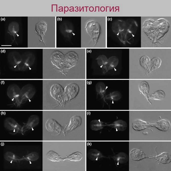
Tubulin localization in Giardia, detected by immunofluorescence microscopy using monoclonal antibodies.
The images show cells at the interphase stage (row a) and at various sequential stages of mitosis (b–d). Using differential interference contrast (DIC) microscopy, tubulin-containing structures (indicated by arrows), axonemes, flagella, and the adhesive disk are clearly visible at all stages of cell division. Scale bar — 5 µm (from: Piva and Benchimol, 2004).
Source: Guadalupe Ortega-Pierres — Giardia and Cryptosporidium, From Molecules to Disease, 2009.
💎 Find even more useful parasitology materials in our LAB-TESTS library. A link with access to the library is automatically sent to you via private message after paying for a VK Donut Labformula subscription.


