

Microscopy of a culture from Dermakit medium, Microsporum canis macroconidia (marked with arrow 1) and Alternaria (arrow 2). Kitten.
👉 The image was kindly provided by Anastasia Kochetkova, Veterinary Clinic “Center”, Rostov-on-Don.
Microscopic examination of dermatophyte culture: differentiating Microsporum canis macroconidia from Alternaria mold in veterinary practice.
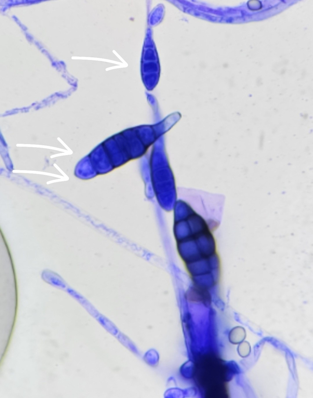
Microscopy of a culture from Dermakit medium, Microsporum canis macroconidia (marked with arrow 1) and Alternaria (arrow 2). Kitten.
👉 The image was kindly provided by Anastasia Kochetkova, Veterinary Clinic “Center”, Rostov-on-Don.

Microscopic findings of hair infection in trichophytosis. Ectothrix-type fungal spore arrangement, absence of Wood's lamp fluorescence, 400x magnification.

Cytological examination of a canine external ear canal smear: morphology of budding yeast-like fungi Malassezia spp. and anucleated epithelium.

A diagnostic cytology exercise: determine which patient clinical history corresponds to the microscopic changes in the smear.