

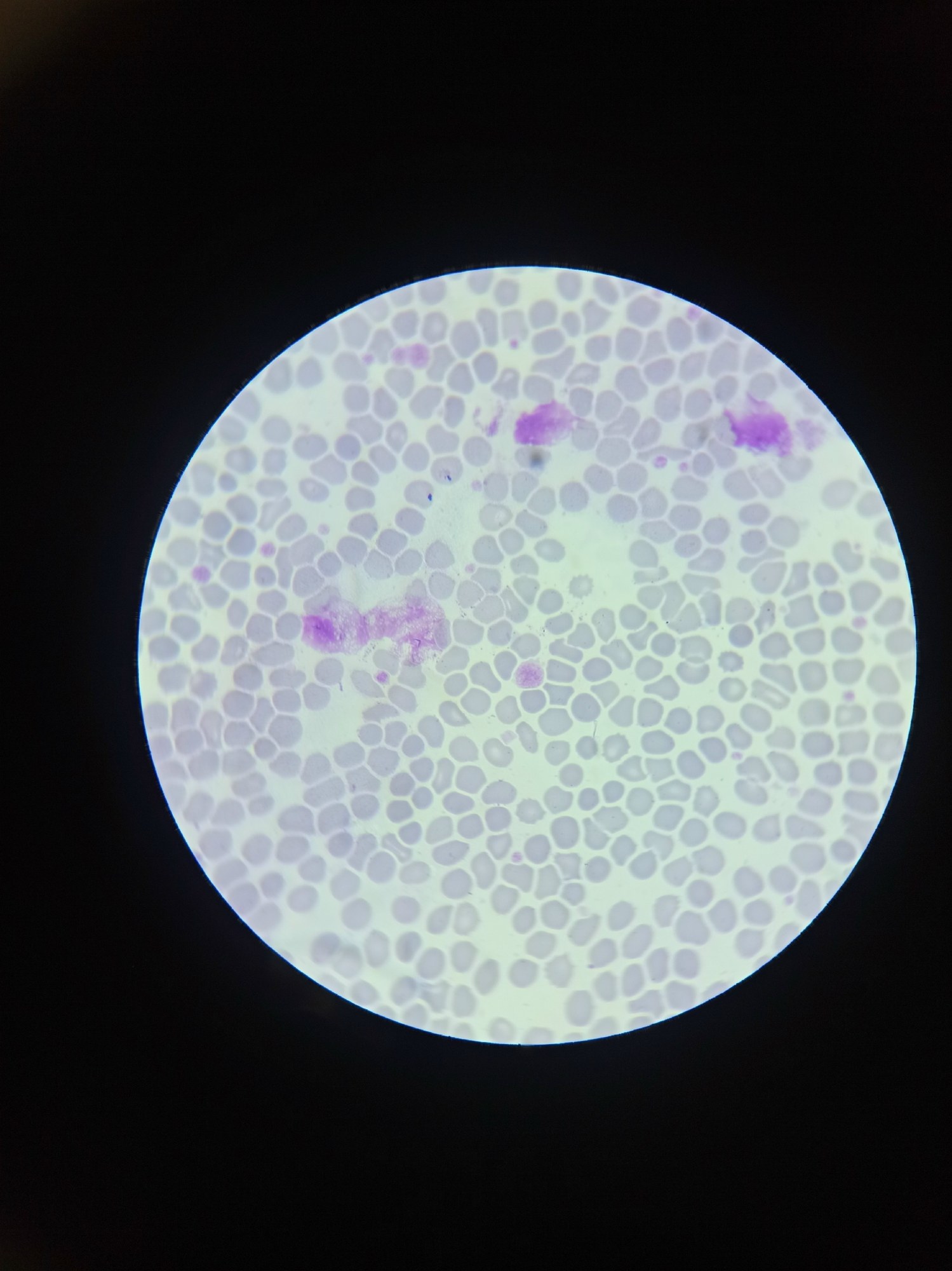
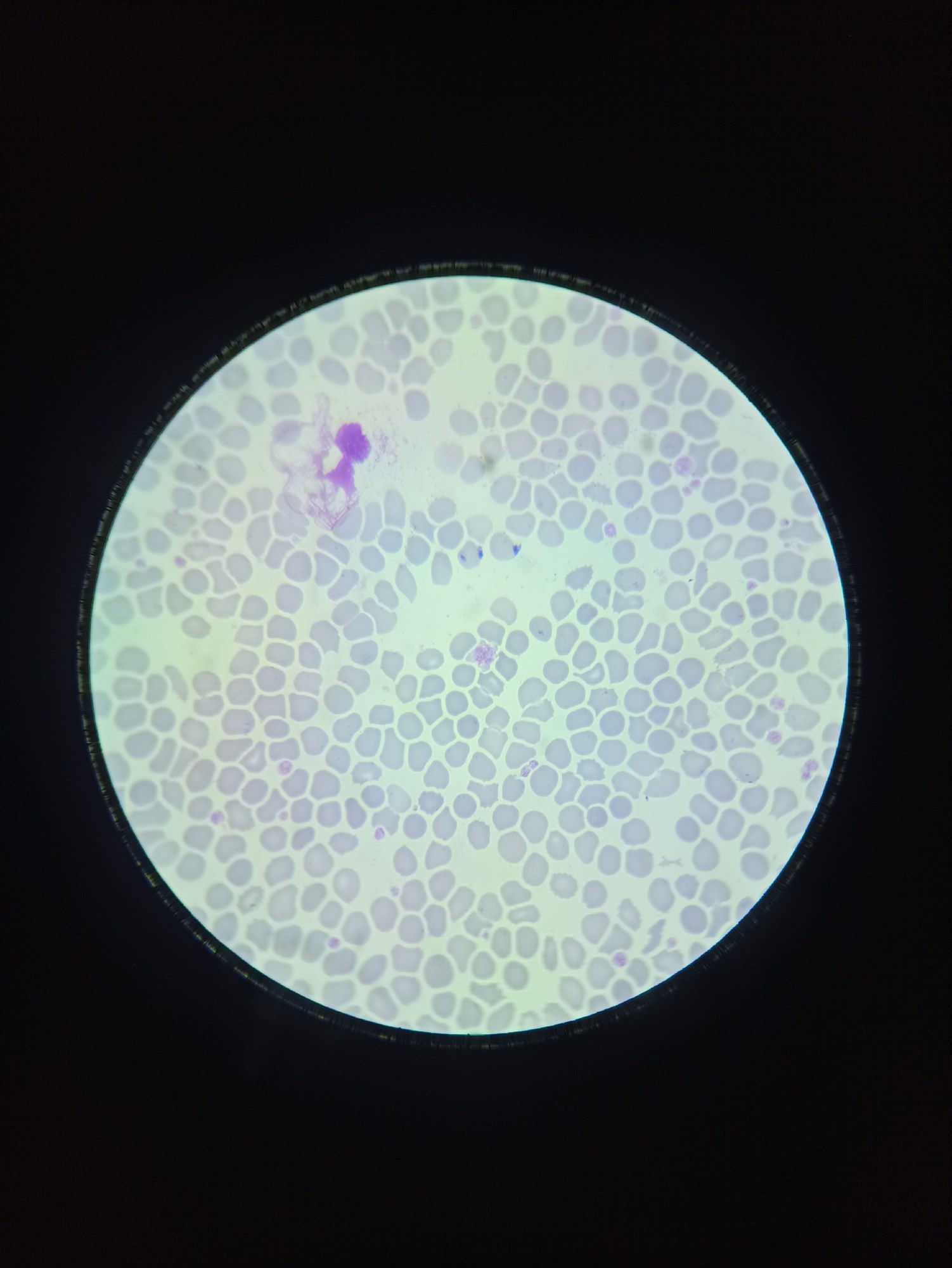
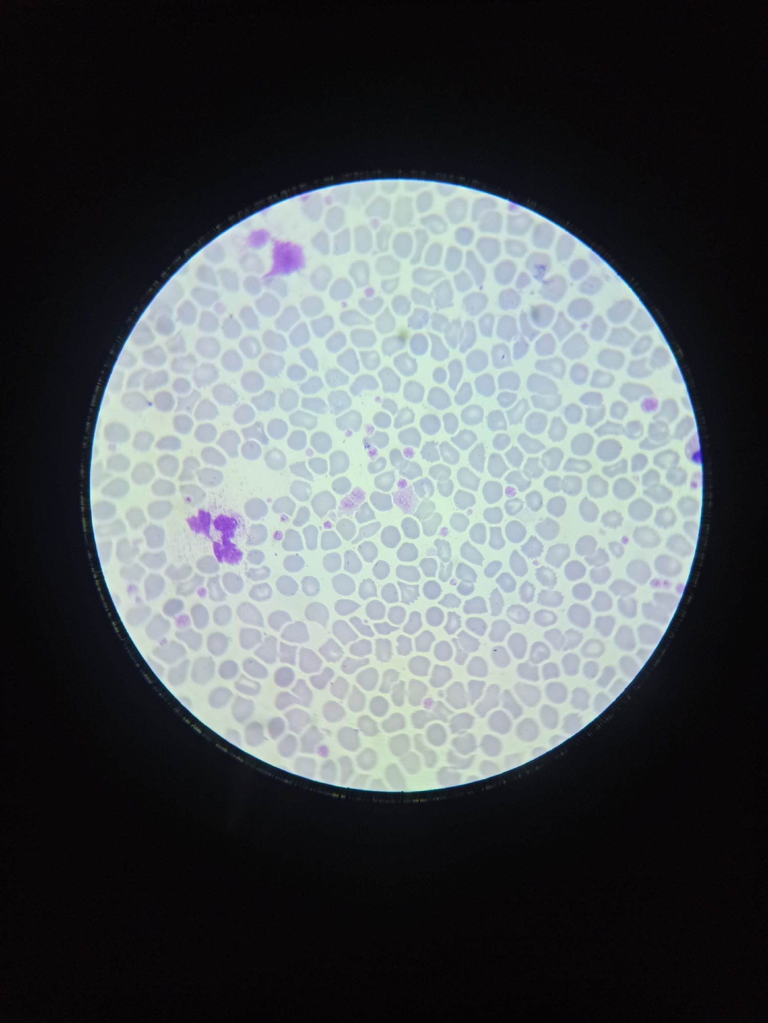
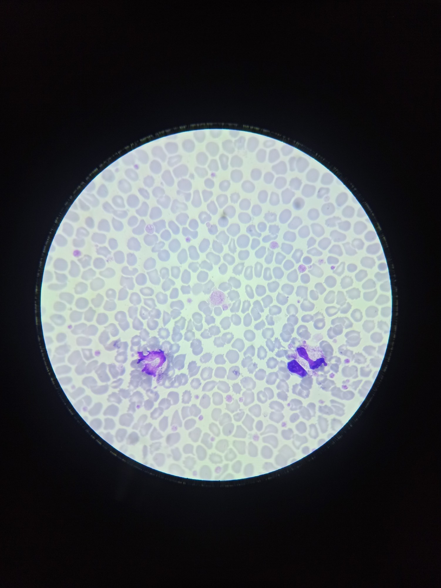
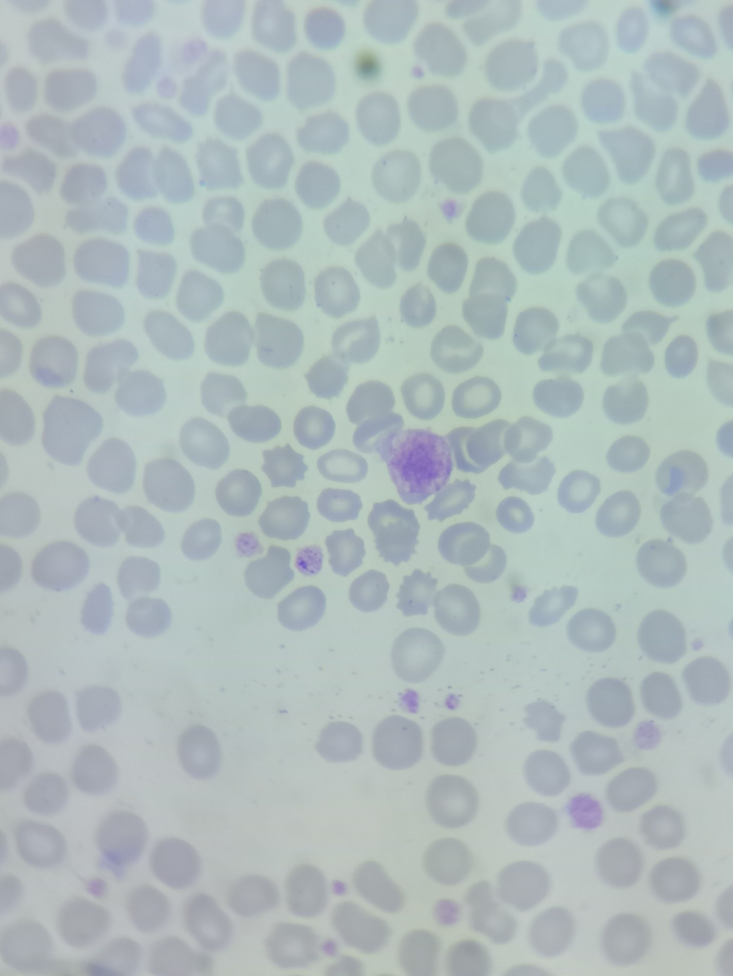
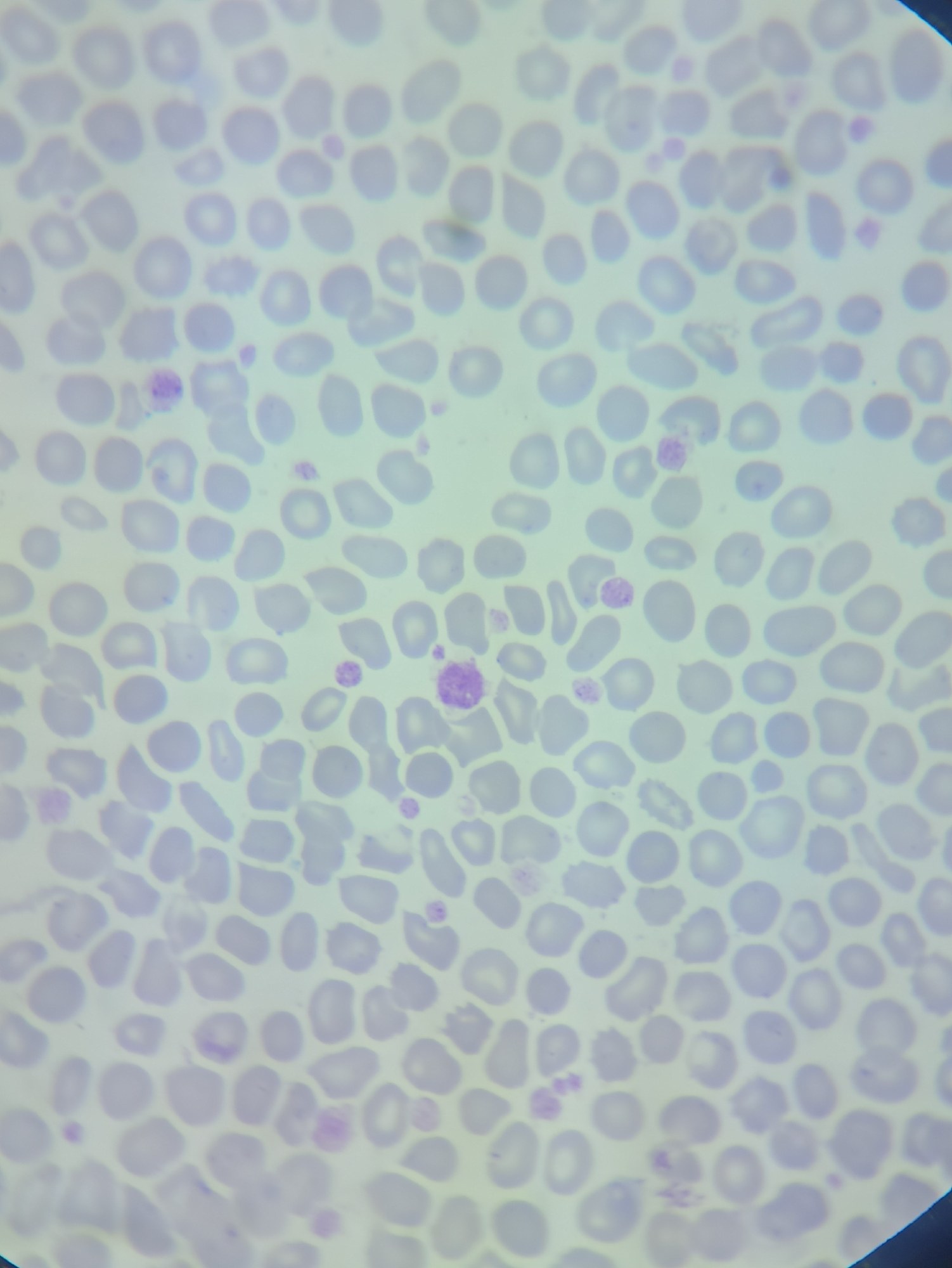
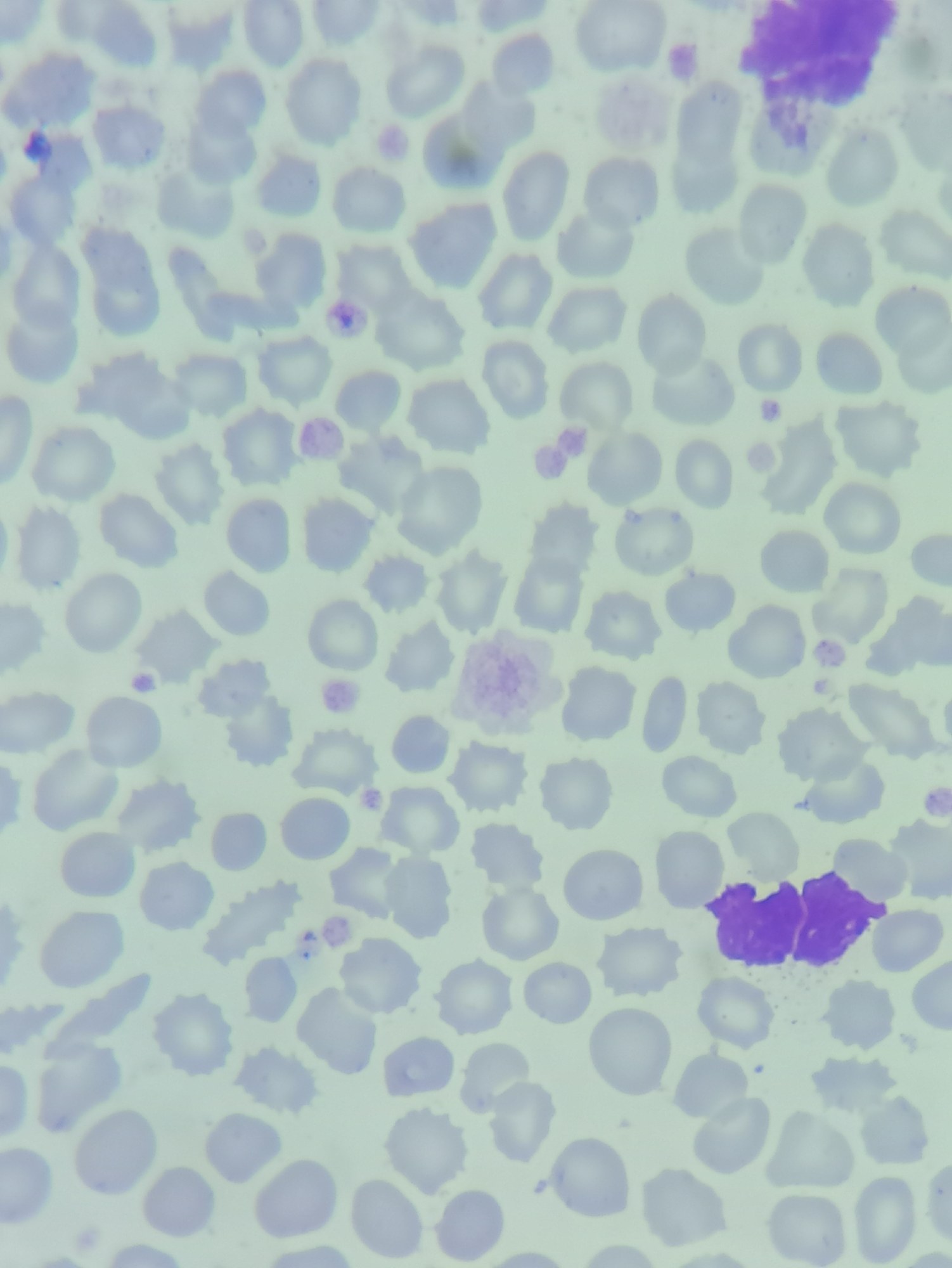
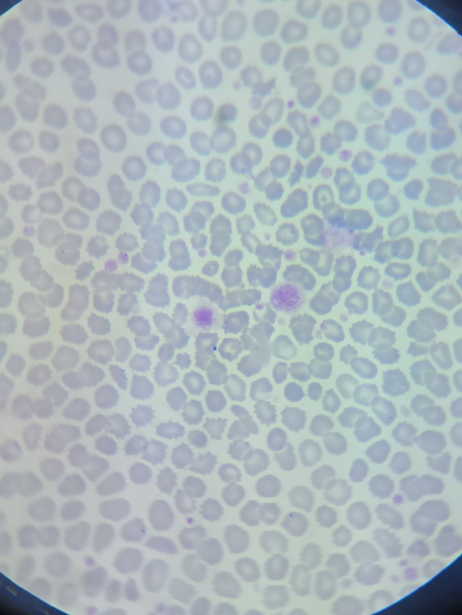
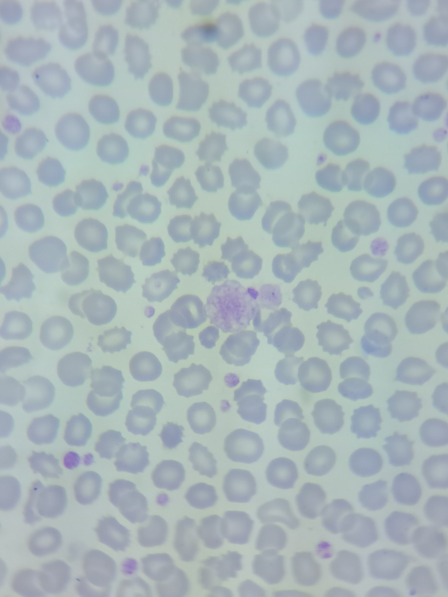
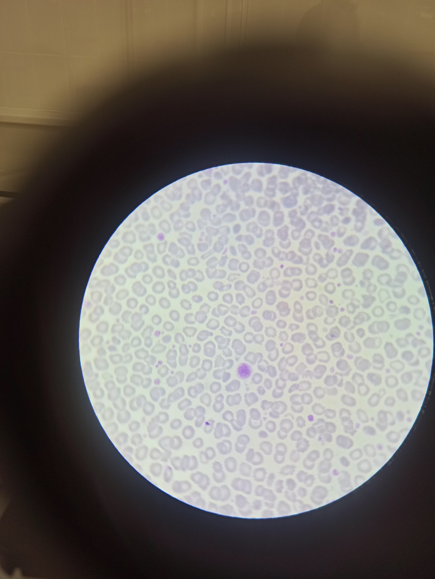
Macrothrombocytes and giant platelets in the peripheral blood of a 74-year-old patient with pancytosis of unknown origin. Complete Blood Count (CBC) parameters: WBC - 29.08 (N 3.5-9.5 × 10⁹/L) RBC - 9.15 (N 3.8-5.8 × 10¹²/L) PLT - 765 (N 125-350 × 10^9/L) (norms are indicated for the analyzer on which the measurement was performed) Macrothrombocytes are abnormally large platelets, whose size can be comparable to that of a normal erythrocyte (7-8 µm); if the platelet size exceeds that of an erythrocyte, they are called giant platelets. Macrothrombocytes and giant platelets appear in peripheral blood in response to inflammation (reactive thrombocytosis), activation (activated platelets, often irregularly shaped, with many protrusions - pseudopods), when taking medications (corticosteroids, cytostatics, GM-CSF), in some leukemias with excessive megakaryocytic proliferation (polycythemia vera, essential thrombocythemia, acute megakaryocytic leukemia), in myelodysplastic syndrome (MDS), primary myelofibrosis (PMF) in early stages, as a manifestation of some hereditary diseases - gray platelet syndrome (deficiency of platelet alpha granules), Bernard-Soulier syndrome, MYH9-RD (May-Hegglin anomaly, Fechtner, Epstein, and Sebastian syndromes). Platelets with increased size pass less efficiently through small capillaries, which can lead to the formation of intravascular microthrombi. In some cases, due to genetic mutations (gray platelet syndrome and Bernard-Soulier syndrome), the adhesive capacity of platelets is impaired, and they lose the ability to adhere to the damaged vessel wall, resulting in impaired platelet-vascular hemostasis and increased bleeding throughout life.
🙏 The material was kindly provided by the medical laboratory technician of Neva Clinic Elisey Zhigulyov, Ulyanovsk


