

🩸 Severe Megaloblastic Anemia in a 48-year-old Patient
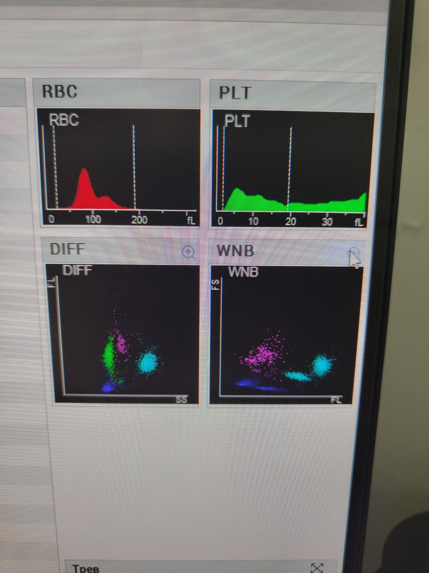
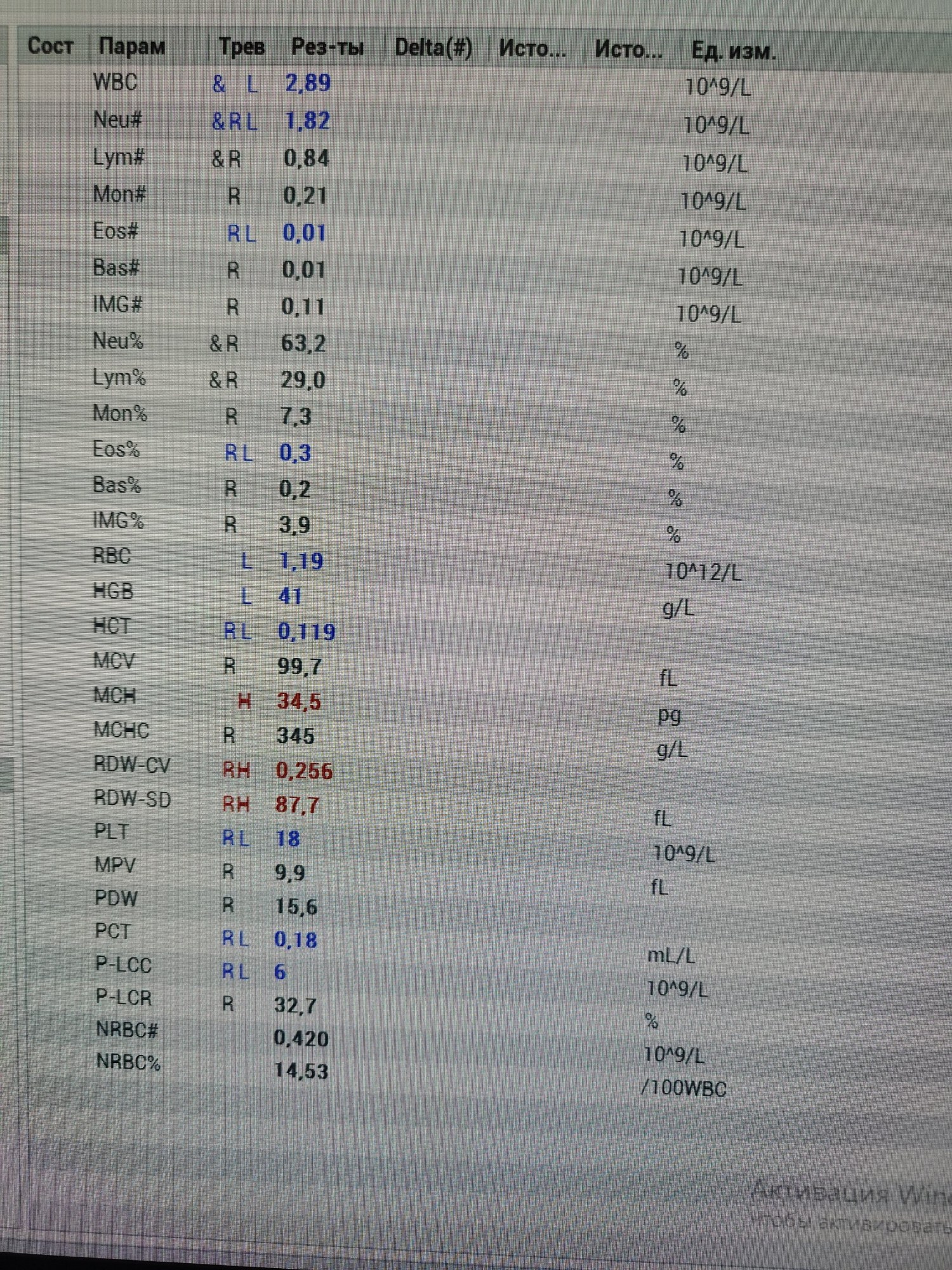
🔬 Peripheral Blood Analysis: UPON ADMISSION: Hemoglobin (Hb): 19 g/L Red Blood Cells (RBC): 0.43 × 10¹²/L White Blood Cells (WBC): 2.5 × 10⁹/L AFTER BLOOD TRANSFUSION: ❗ CBC after blood transfusion is shown in the image.
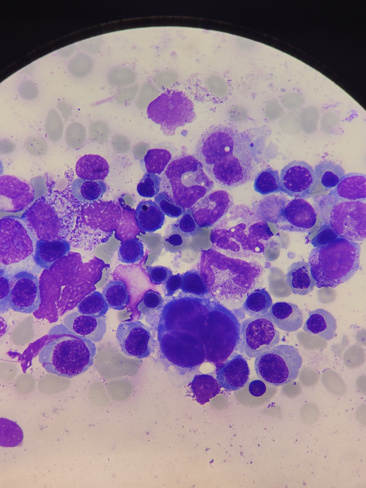
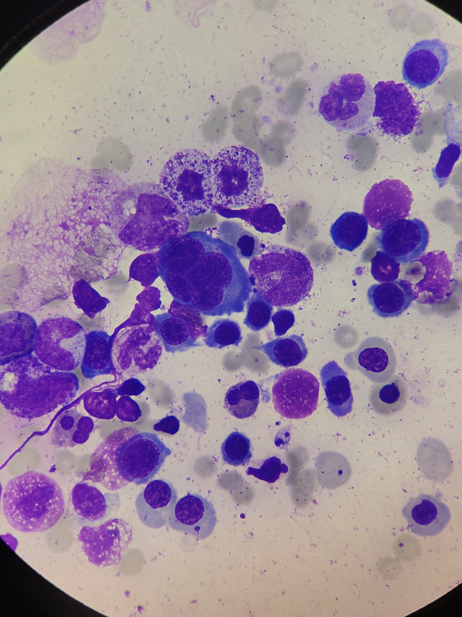
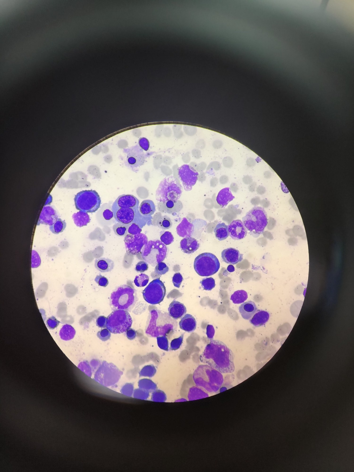
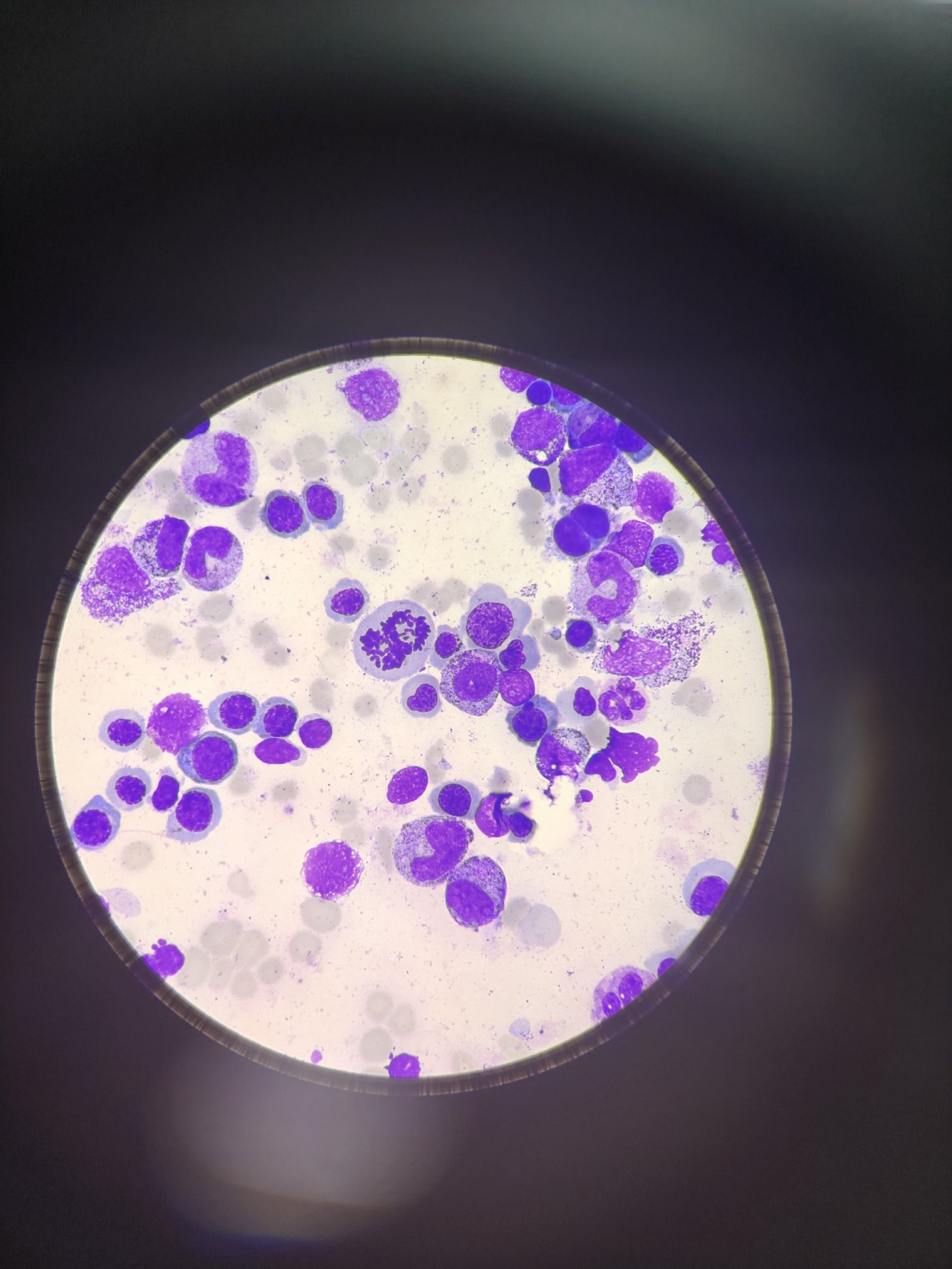
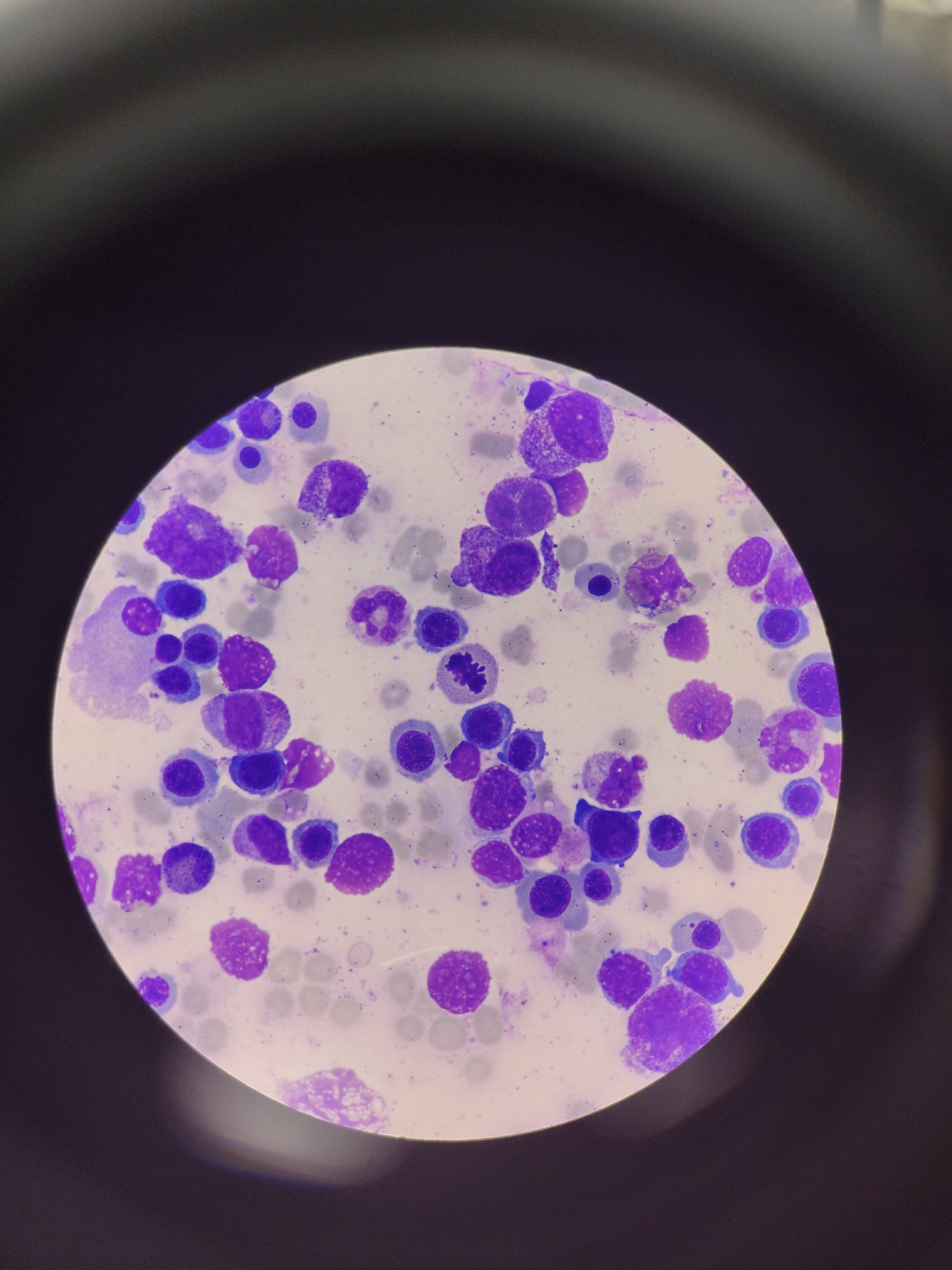
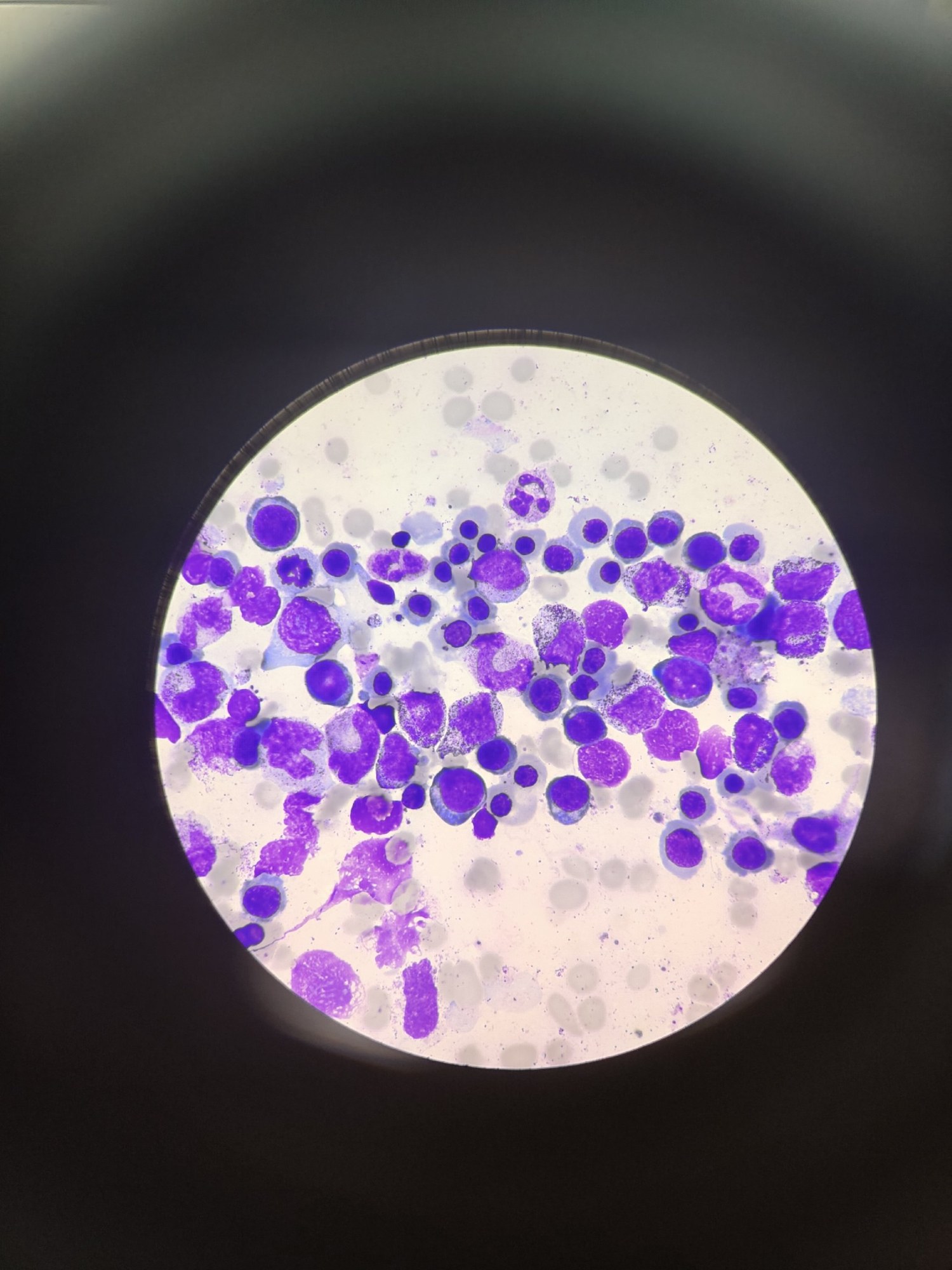
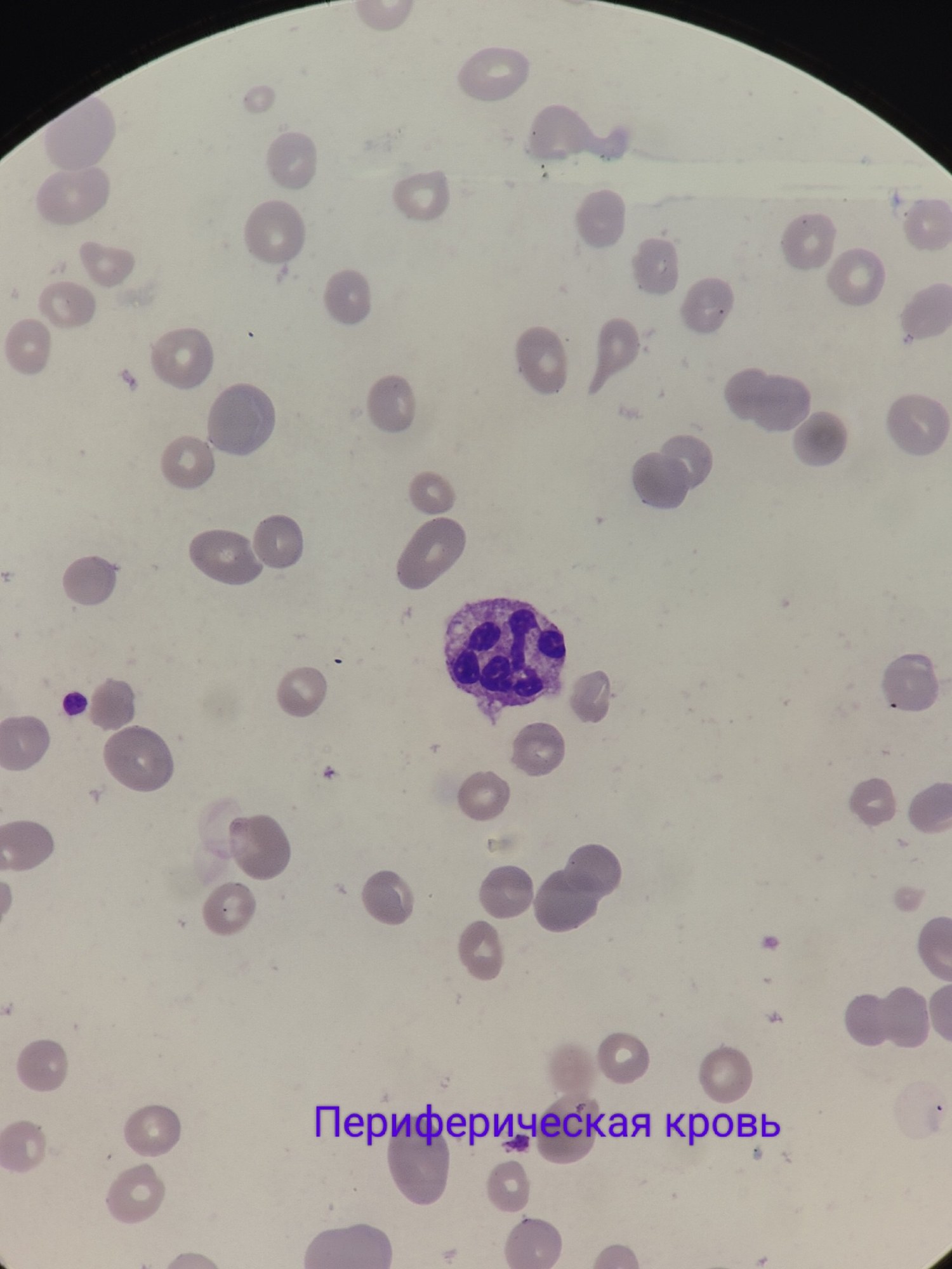
🦴 MYELOGRAM The bone marrow shows a pronounced predominance of megaloblastic hematopoiesis, characteristic of this type of anemia. Key morphological changes: ✅ Megaloblasts constitute up to 70% of cells. Bi- and trinucleated forms are found. ✅ Karyorrhexis (nuclear fragmentation) is observed in erythroblasts. ✅ Giant myelocytes and band neutrophils are present. ✅ Hypersegmented neutrophils are characteristic. 📋 Conclusions The morphological findings from the bone marrow and peripheral blood confirm the diagnosis of megaloblastic anemia, likely caused by vitamin B12 or folic acid deficiency.
💡 To clarify the diagnosis, it is recommended to: 📊 Determine blood levels of vitamin B12 and folate. 🧪 Perform a test for intrinsic factor antibodies. 📎 Assess the state of the gastrointestinal tract (e.g., gastroscopy) to rule out malabsorption of vitamins.
🙏 Clinical case kindly provided by biologist Anna Feditskaya, Luhansk.


