

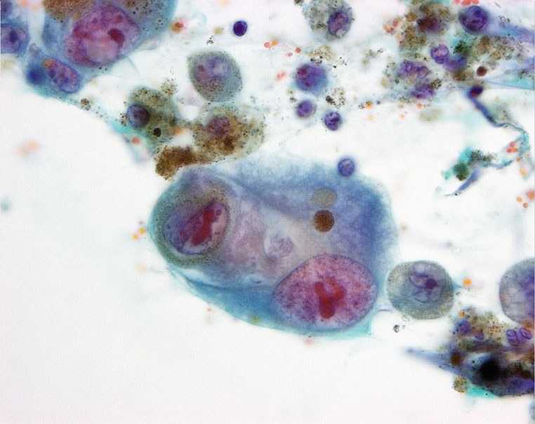
Metastatic melanoma in ascitic fluid. Tumor cells are discretely arranged, with abundant cytoplasm and eccentric oval nuclei containing prominent nucleoli. Characteristic melanin pigment (brown granules) is visible in the cytoplasm. The pigment is also phagocytosed by surrounding macrophages. Location: ascitic fluid.
Source: M. Sheaff, N. Singh – Cytopathology: An Introduction (2013).


