

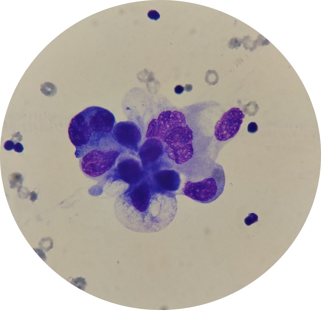
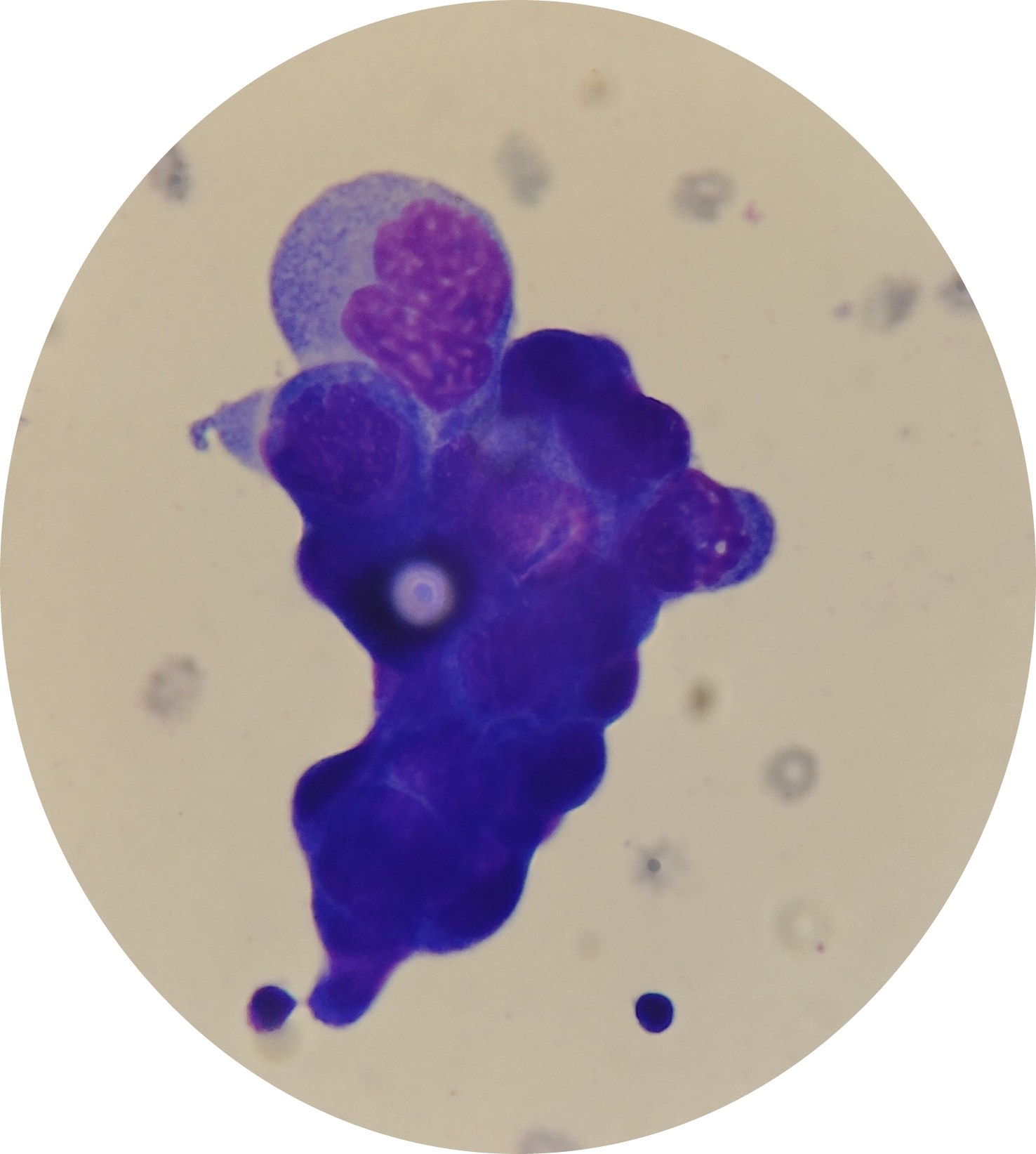
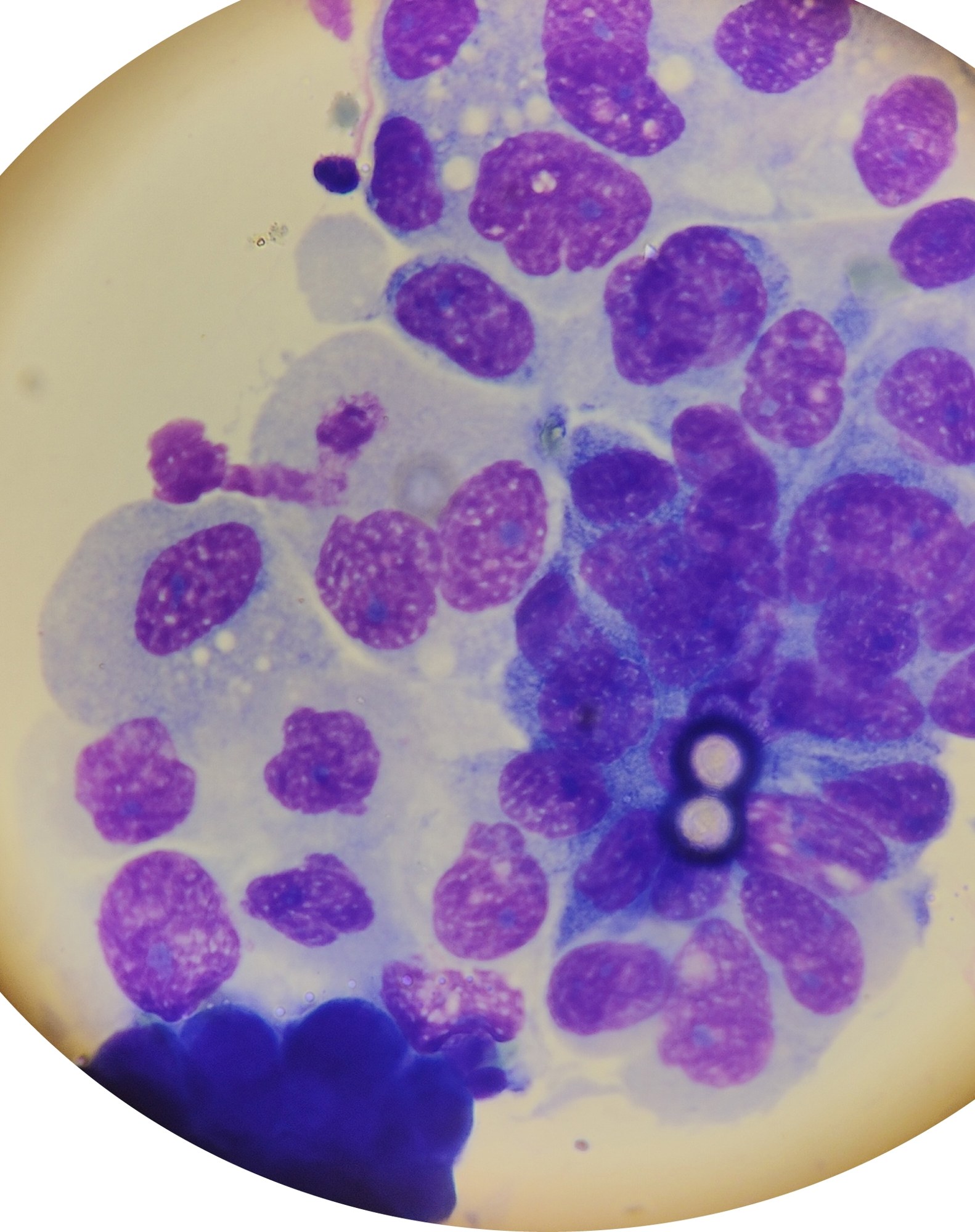
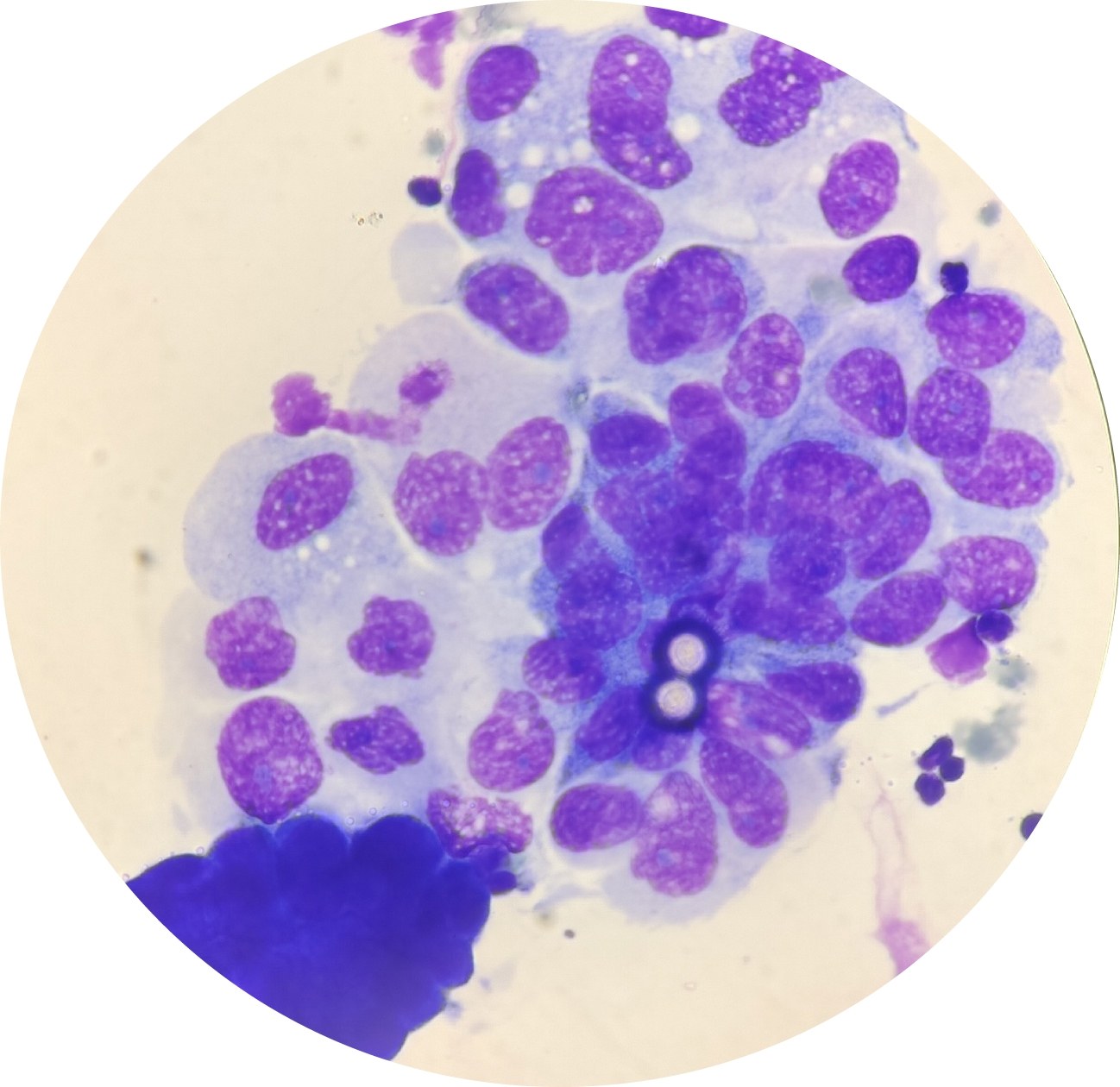
💧Pleural fluid examination in a 79-year-old male. 👉 Diagnosis: Tumor formation in the upper lobe of the right lung.
🔴 Hemorrhagic exudate, protein 27.2 g/l. Color: Hemorrhagic before centrifugation; after centrifugation, the supernatant has a xanthochromic tint.
🔬 Sediment microscopy: ✅Erythrocytes – altered and unaltered. ✅ Leukocytes – neutrophils and lymphocytes in equal proportions, up to 50 per field of view. ✅ Macrophages – histiocytic cells, present individually and in small groups. ✅ Mesothelial cells – moderate reactive changes. ❗ Metastatic glandular carcinoma cells (MTS) detected.
🙏 Photos and description kindly provided by biologist Anna Feditskaya, Luhansk.


