

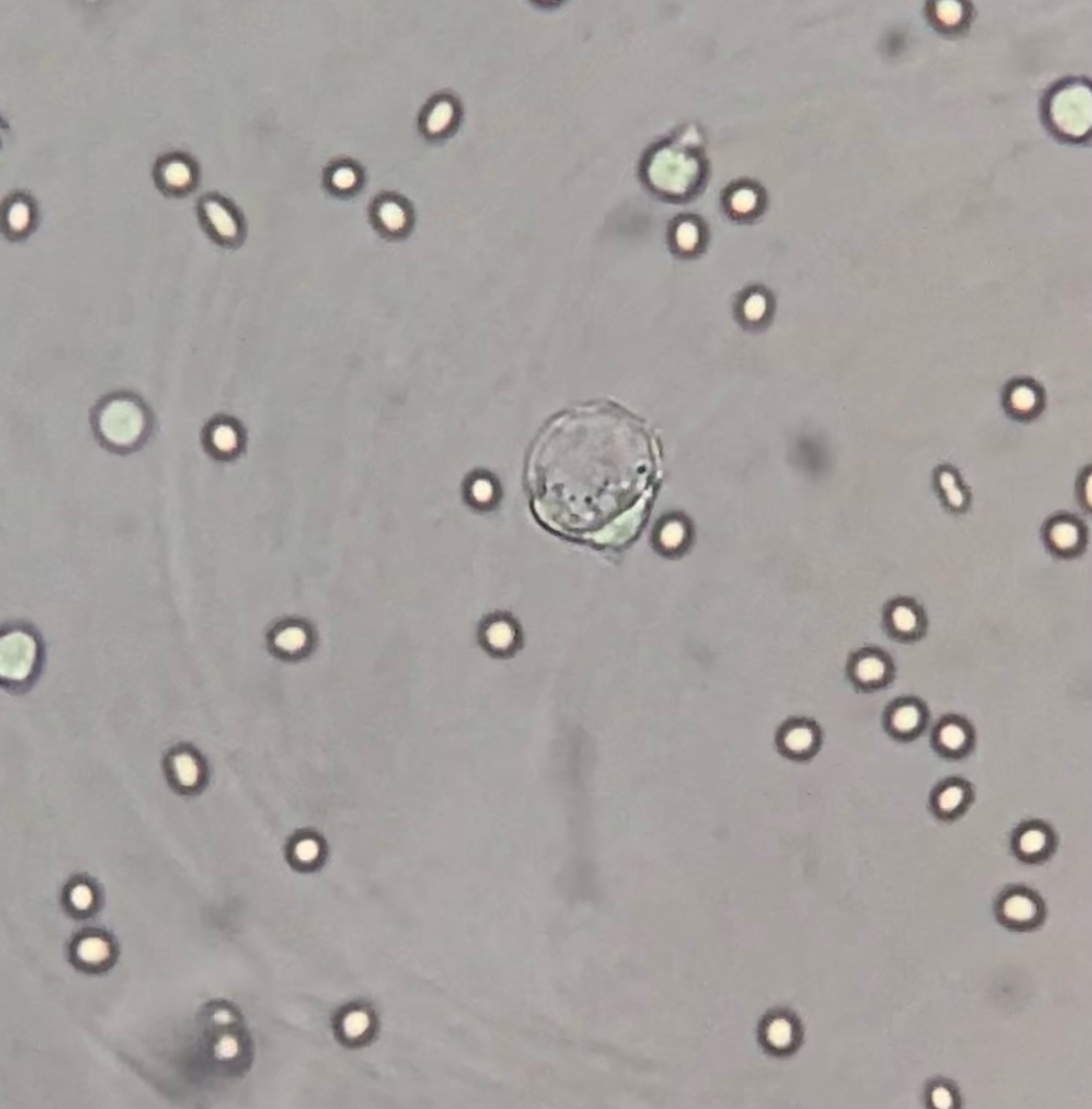
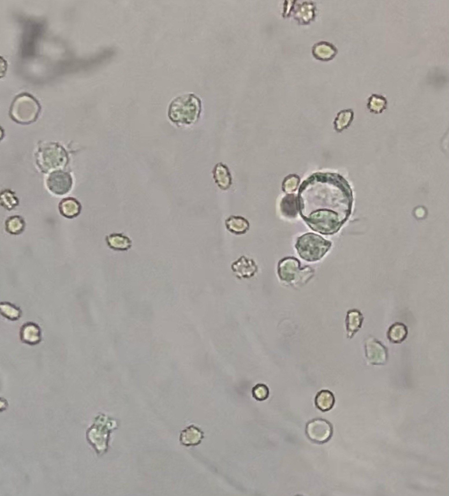
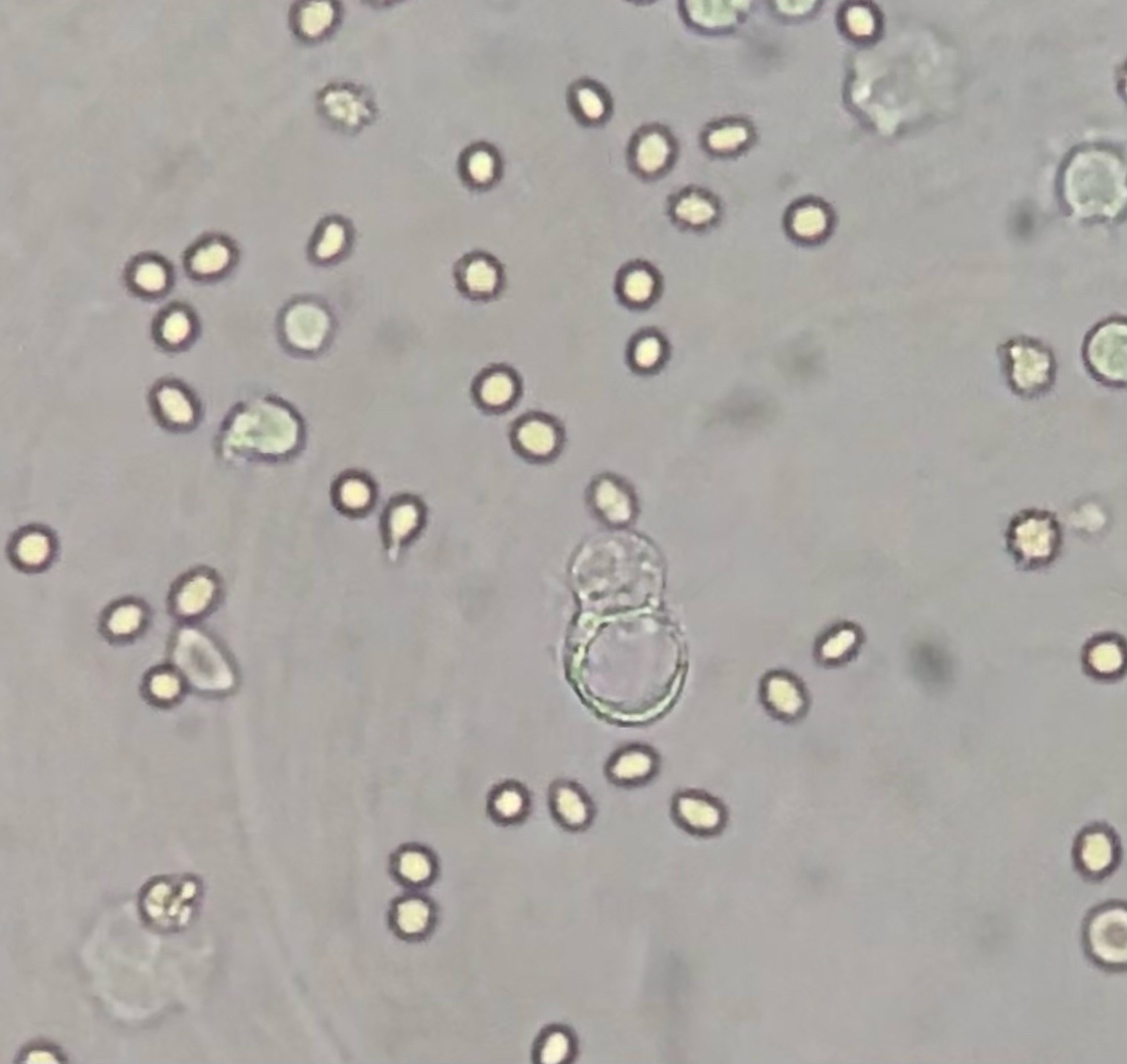
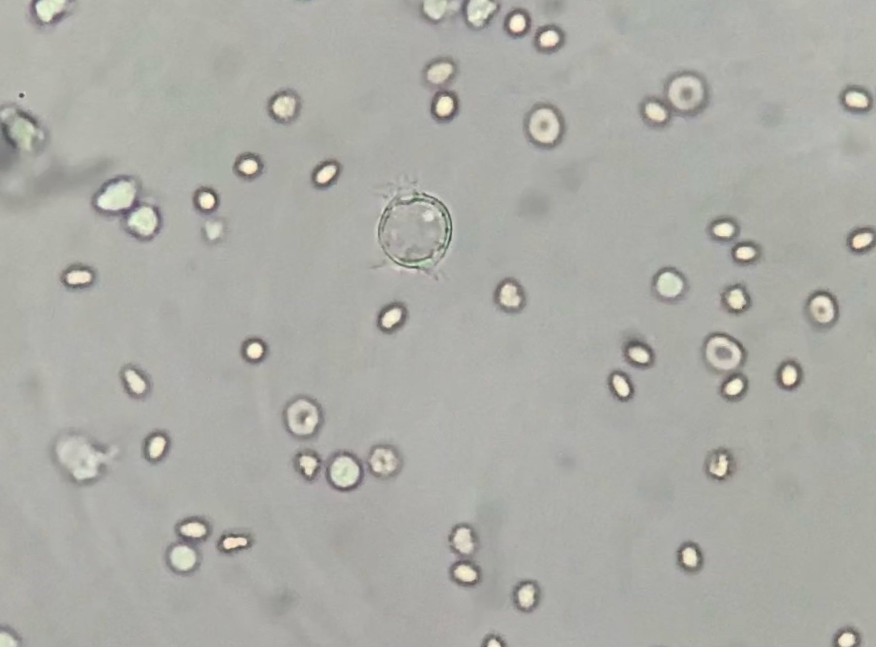
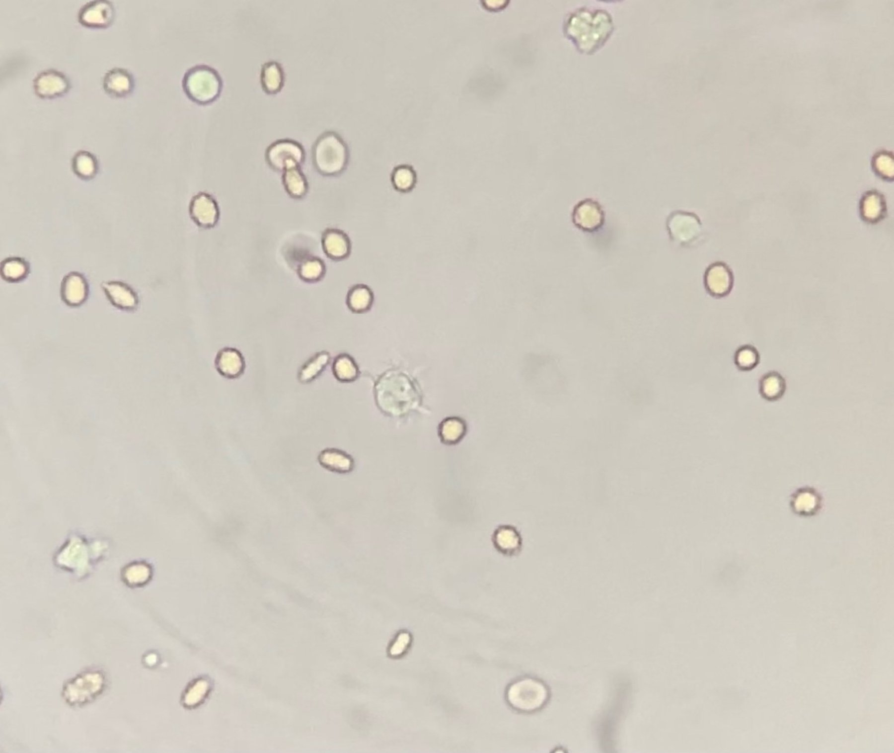
🔬 Ascitic fluid. Native specimen. Mesothelium against a background of erythrocytes and leukocytes. Magnification 400x.
Reference information: Mesothelium is a single layer of squamous epithelium of serous membranes lining body cavities (peritoneum, pleura, pericardium) in vertebrate animals and humans, formed from the mesoderm. It consists of flat polygonal cells with tightly closed edges. In mammals and humans, the outer surface of mesothelial cells is covered with microvilli, which are involved in the absorption and secretion of cavity fluid by these cells.
🙏 Material kindly provided by KDL biologist Yulia Guseva, GBUZ Belorechenskaya Central District Hospital.


