

Fungal mycelium. Microscopy of a foot nail specimen. Magnification x400. ✅ Pre-treatment of biomaterial with alkali in vitro.
🙏 Image kindly provided by Clinical Laboratory Diagnostics Physician, Anna Ivanova, Moscow
Microscopic image of fungal mycelium in a foot nail scraping, suspected onychomycosis. Features of slide preparation with alkali treatment.
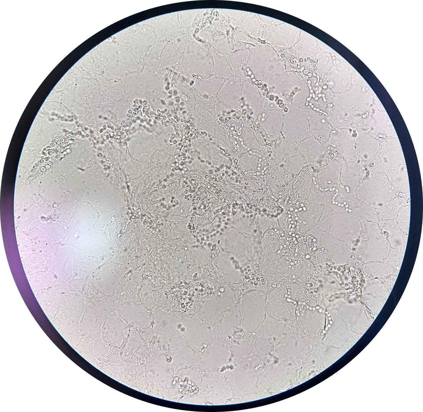
Fungal mycelium. Microscopy of a foot nail specimen. Magnification x400. ✅ Pre-treatment of biomaterial with alkali in vitro.
🙏 Image kindly provided by Clinical Laboratory Diagnostics Physician, Anna Ivanova, Moscow

Examples of skin and nail scraping microscopy in mycoses: visualization of mycelial threads, arthrospore chains, and pathogenic fungal elements in native preparations.

Microscopic examination of a nail plate scraping for suspected onychomycosis. Detection of branching fungal mycelial threads in a native preparation.

Microscopic view of arthroconidia and sporulated mycelium in 20% KOH preparations from skin and nail mycoses caused by dermatophytes.