

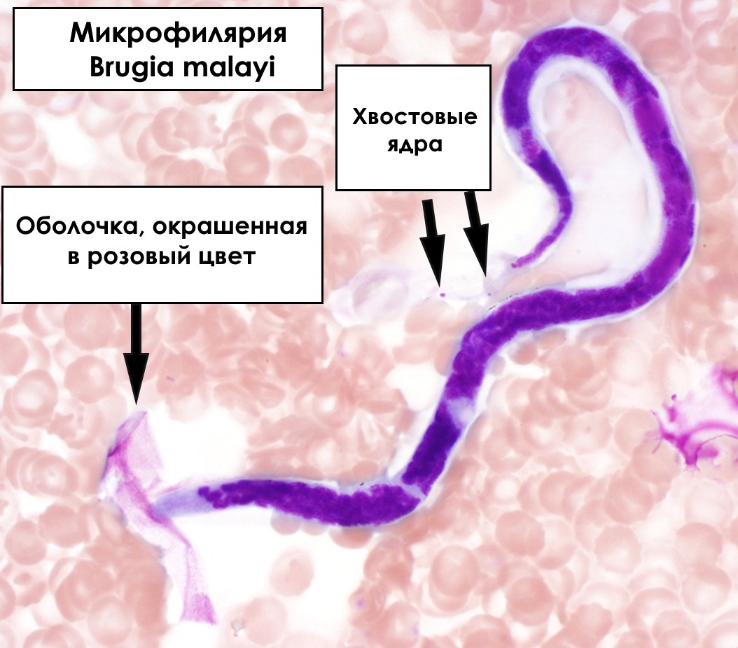
Answer to the parasitology problem: Brugia malayi microfilariae
These microfilariae have a clearly visible sheath, which immediately helps narrow down the possible options to three: Wuchereria bancrofti, Loa loa, and members of the genus Brugia. Further differences are determined by the structure of the head, tail, and the arrangement of nuclei in the tail region.
In this case, two nuclei are visible in the tail, located far apart — closer to the tip. This pattern is characteristic of Brugia malayi. Staining also helps: the densely stained pink sheath distinguishes B. malayi from B. timori.


