

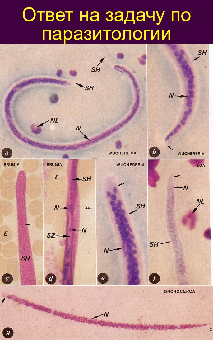
🐍 CORRECT ANSWER TO THE PARASITOLOGY EXERCISE. Photographs of human blood microfilariae (a) Wuchereria bancrofti, entire microfilaria. (b) W. bancrofti: posterior end of microfilaria; nuclei do not reach the very tip of the tail (arrow). (c) Brugia malayi: sheath (SH) is intensely stained and appears granular (double arrow). (d) Brugia malayi: posterior pole; one nucleus is located precisely at the terminal pole (double arrow), and another is some distance from it. (e) W. bancrofti: anterior end; the anucleated region is equal in size to the width of the larva. (f) Loa loa: although the worm is stained, the staining is weak; therefore, the nuclei are difficult to distinguish, but they are also found at the anterior pole. (g) Onchocerca volvulus: entire microfilaria, obtained from a lymphatic vessel. The anterior and posterior regions are clearly free of nuclei.
Labels: E — erythrocyte, N — nucleus, NL — leukocyte nucleus, SH — sheath, SZ — tail. 💎 Find more useful parasitology materials in our LAB-TESTS library. The link with access to the library is automatically sent to you via private message after paying for a VK donut Labformula subscription.


