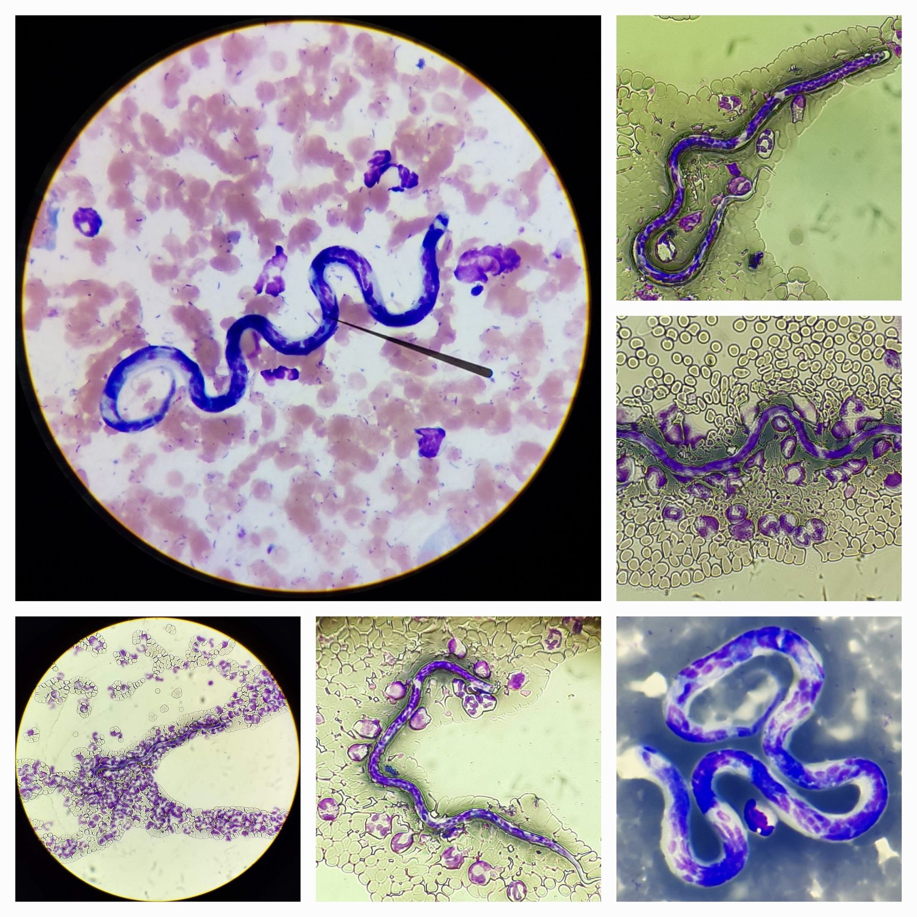
🔬 Microfilaremia. ✅ In our region, cutaneous dirofilariasis, transmitted by mosquitoes, is common in dogs. Larvae (microfilariae) can often be seen in large breeds during blood or even urine examination in cases of hematuria, or in highly hemodiluted tumor aspirates, as an incidental finding, without clinical manifestations. ✅ The cardiac form of dirofilariasis is of greater importance, as it can lead to the death of the dog. However, we more often suspect it in transit animals. Morphological differentiation of microfilariae (larvae) of different forms in the blood is not possible with standard staining. Special, prolonged staining for acid phosphatase is required. By the way, the internal contents of microfilariae are Wolbachia, also living organisms, commensals. 👉 The clinical case was kindly provided by - Olga Belozerova, clinical pathologist, head of the laboratory at the veterinary clinic “Noah’s Ark”, Kirov.
Microfilariae in Blood Smear: Diagnosing Dirofilariasis
Detection of microfilariae in peripheral blood smears in cutaneous and cardiac dirofilariasis. Features of differential morphological diagnosis of larvae.