

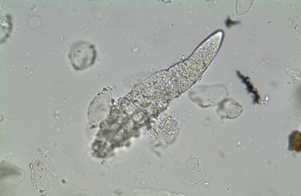
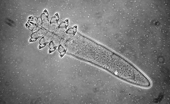
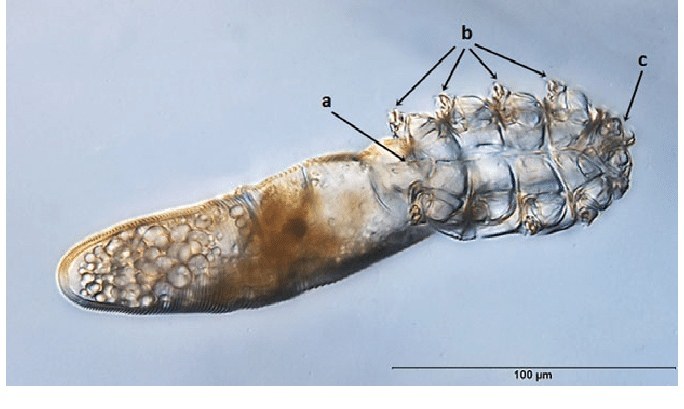
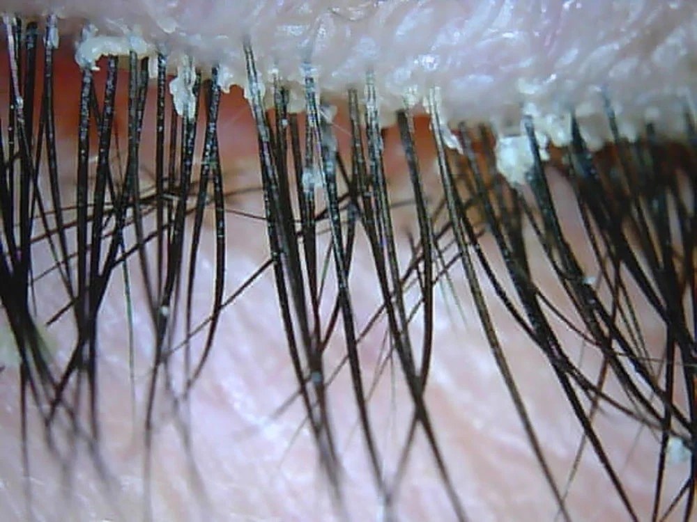
🔬Demodex. Microscopy. ⚠Noticed this symptom: “Eyelashes sticking together in the morning”? ✅Microscopic examination of eyelashes or skin scraping is used to identify the Demodex parasite. ✅Infection with Demodex folliculorum more often leads to itchy, slightly flaky red areas. If Demodex brevis is the main causative agent, the appearance of papules (“pimples”) covered with gray scales comes to the forefront. Sometimes, instead of these, vesicles with clear or purulent contents, surrounded by a red inflammatory rim, are found on the skin.
💎Look for a video recording of Demodex mite microscopy in our LAB-TESTS library. The link with access to the library is automatically sent to you via private message after paying for a VK donut Labformula subscription.


