

🔬 Cerebrospinal Fluid Analysis: Meningitis Diagnosis.
A 3-year-old child was admitted with complaints of headache 🤕, fever 🌡, chills ❄, and a high temperature spike. Clinically, nuchal rigidity 🤷♂, difficulty flexing the neck, increased sensitivity to light 💡 and sounds 🔊, as well as nausea 🤢 and vomiting 🤮 accompanying the headache were noted. A hemorrhagic rash was also found on the child’s skin.
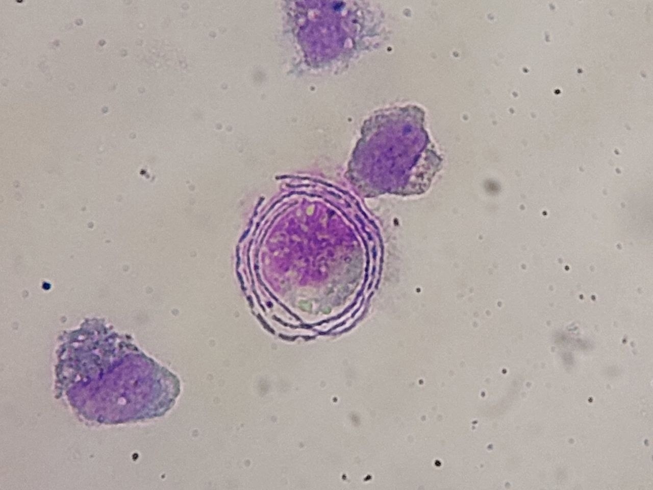
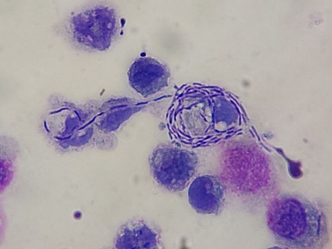
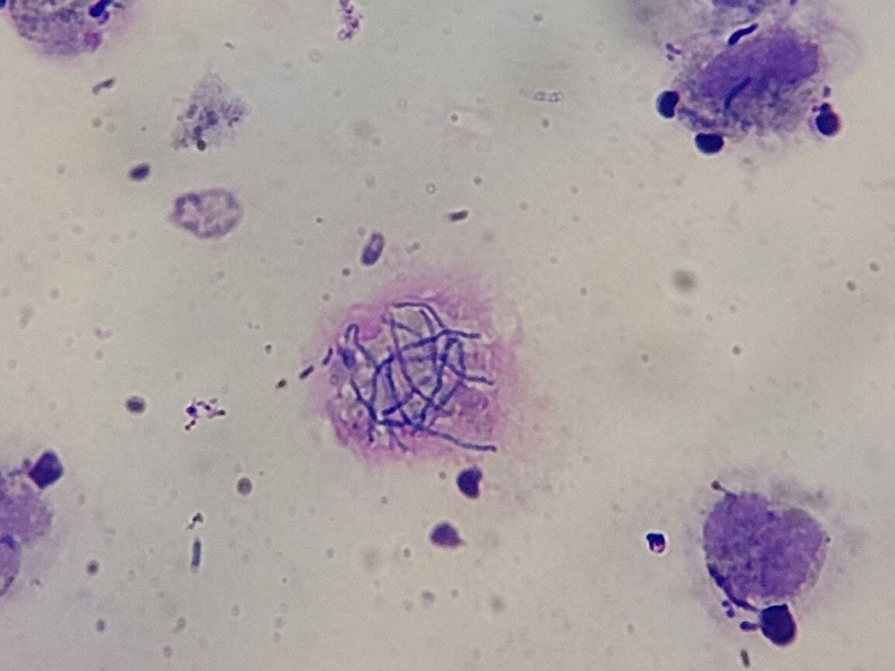
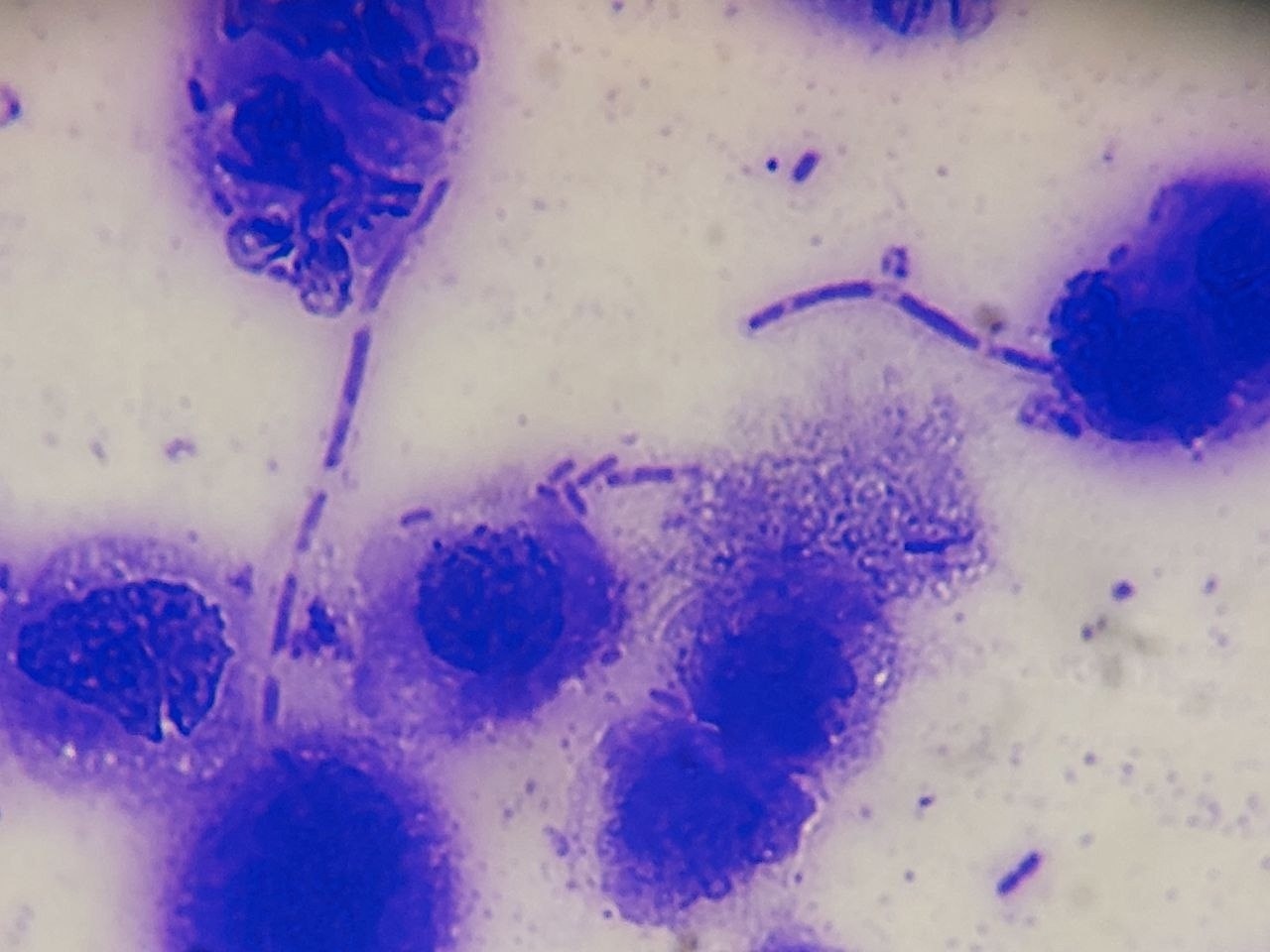
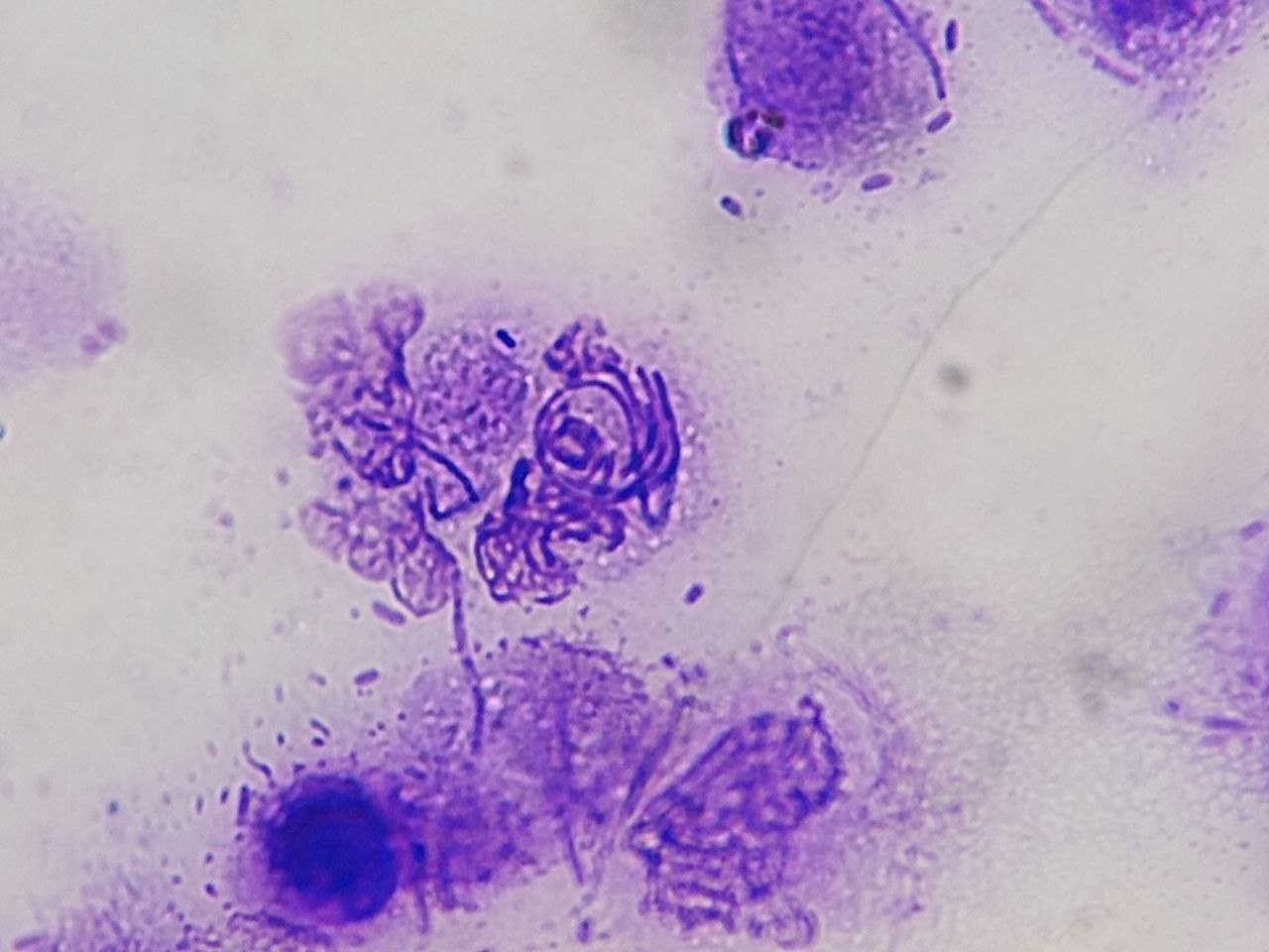
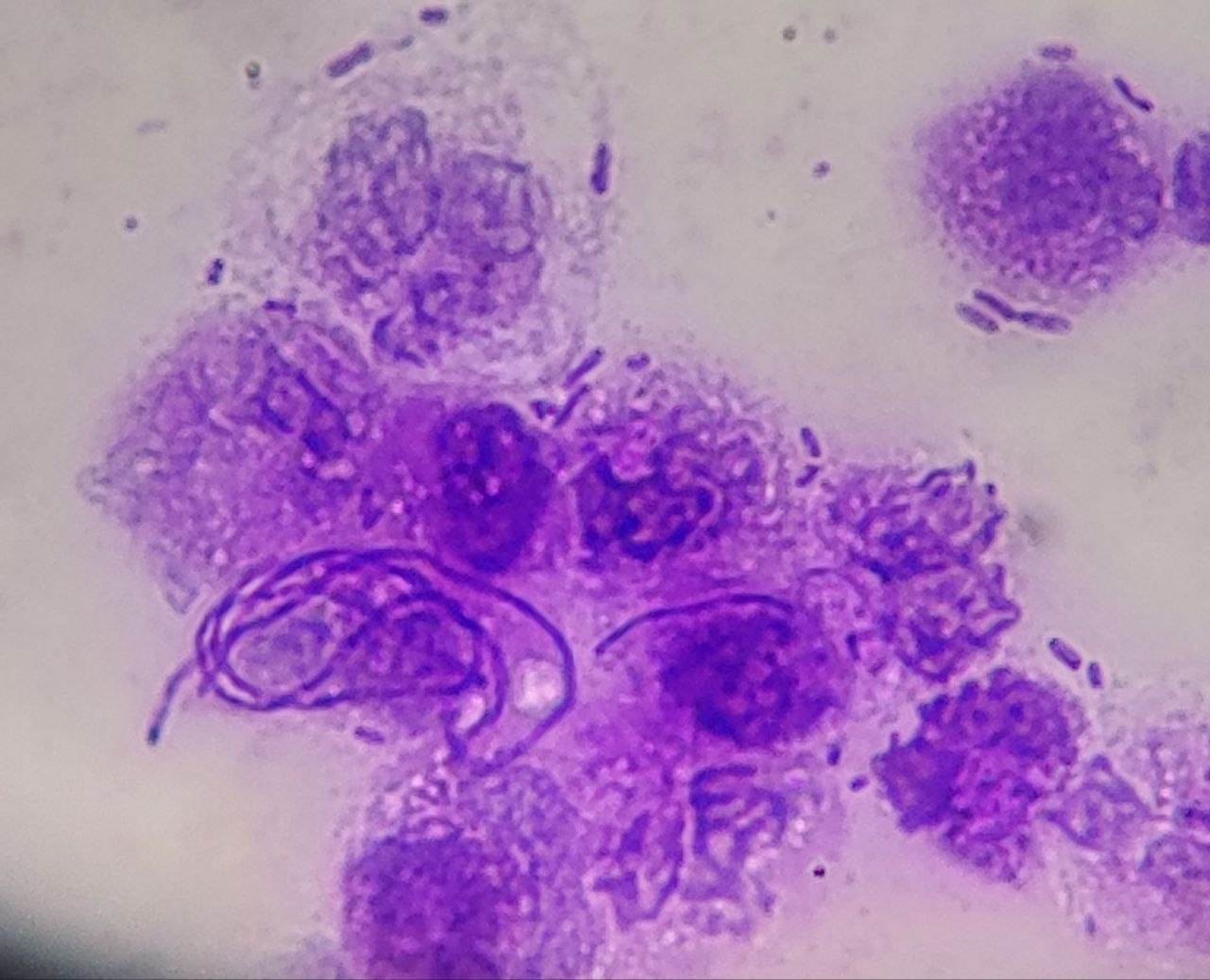
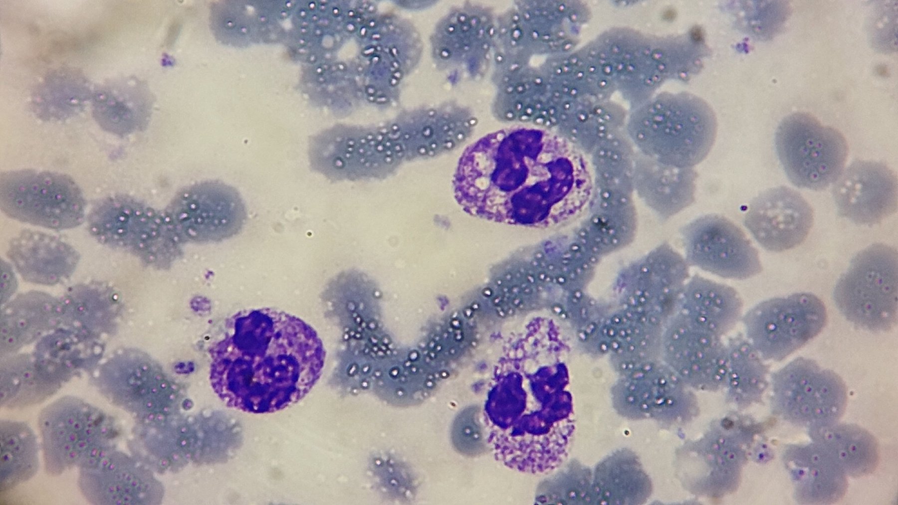
To diagnose suspected meningitis, a lumbar puncture 💉 was performed, followed by cerebrospinal fluid analysis. The cerebrospinal fluid was turbid, with pronounced neutrophilic and lymphocytic pleocytosis. The protein level was 100 mg/dL, indicating an inflammatory process in the central nervous system 🧠. Microscopic examination with methylene violet staining revealed the presence of specific pathogens (images: 1-6 below).
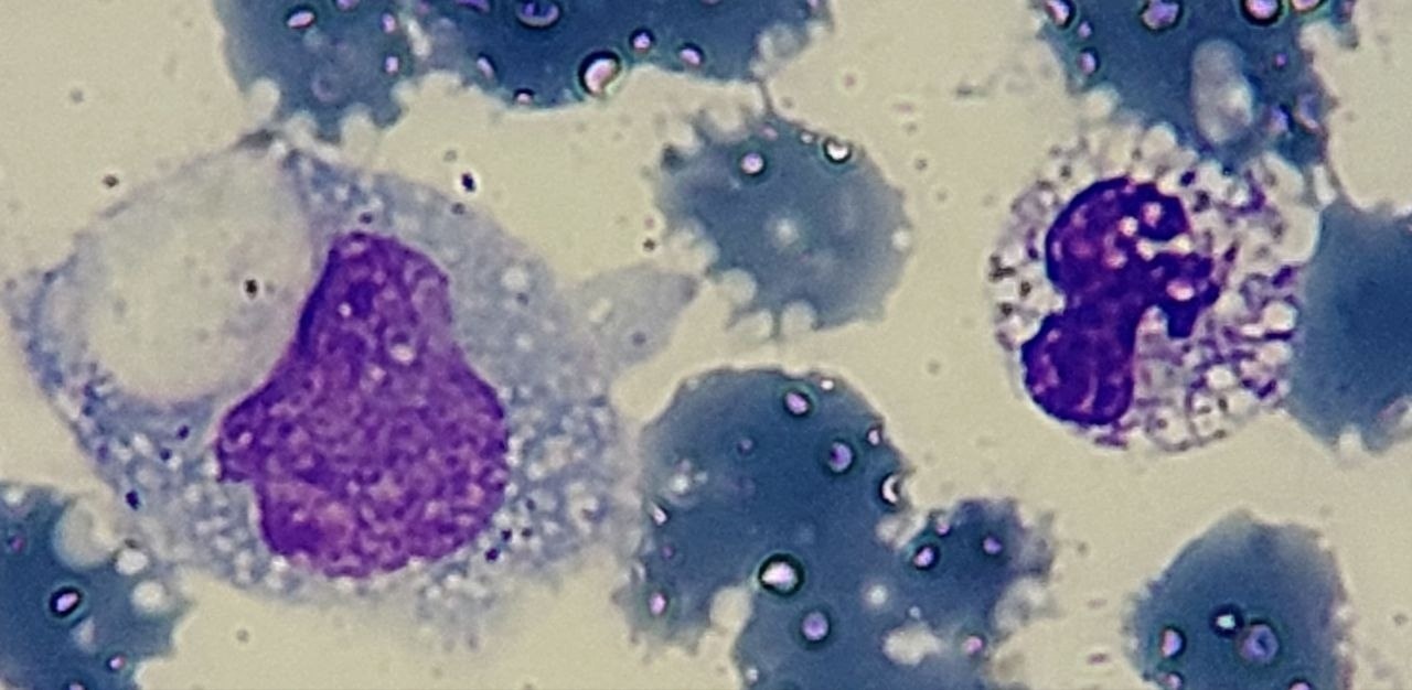
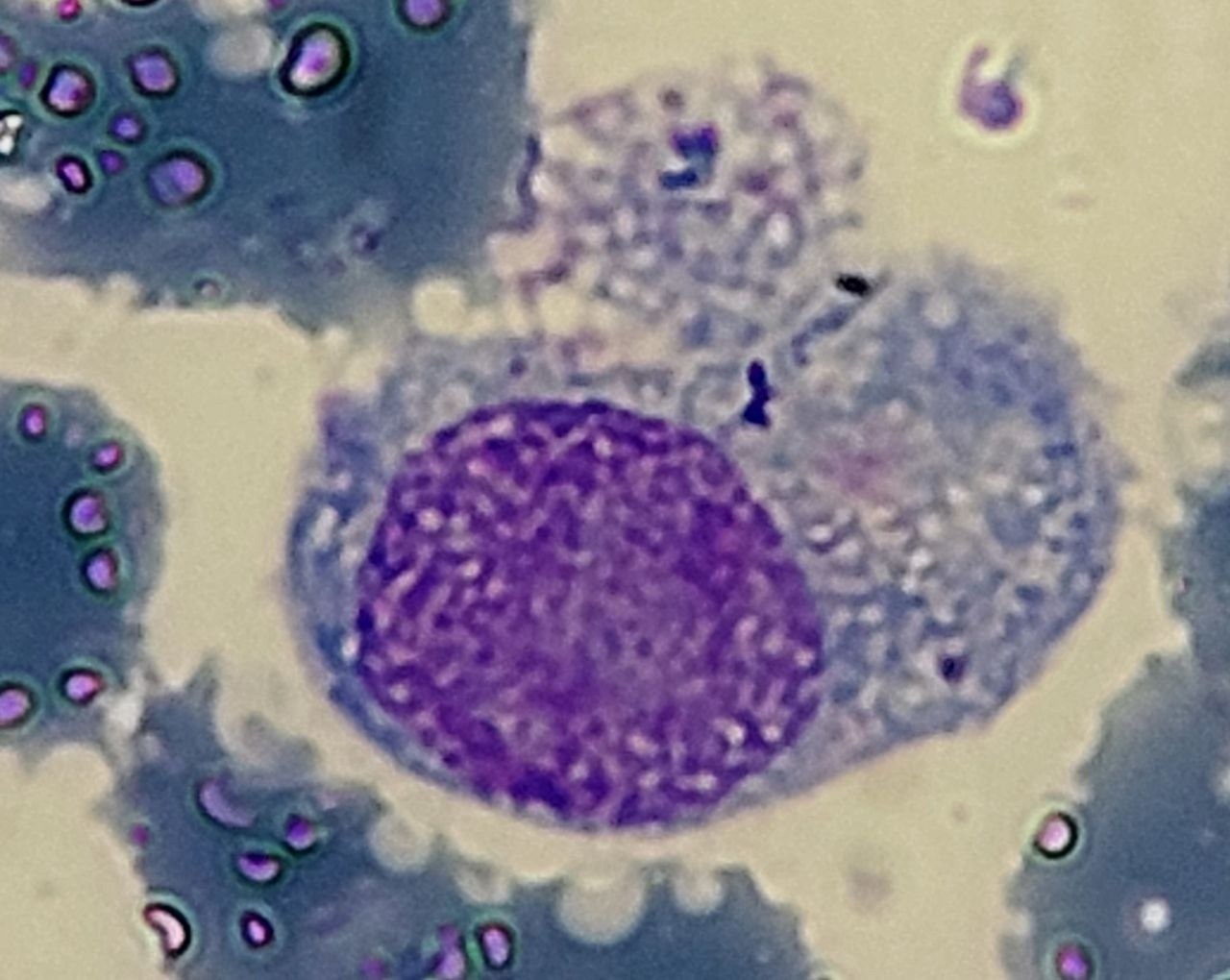
🩸 Complete Blood Count: Hemoglobin: 78 g/L (severe anemia); Leukocytes: 25×10⁹/L (leukocytosis) 🔬; Platelets: 350×10⁹/L (within normal limits). Cell morphology indicates pronounced neutrophilia with vacuolization and toxic granulation in neutrophils and monocytes (photos 7,8,9).
❓ What infectious agent could be the likely pathogen in this patient?
🙏 Clinical case kindly provided by Zebiniso Kholmuradova, morphologist, 🇺🇿 Uzbekistan, Kashkadarya Multidisciplinary Children’s Medical Center.


