

Autumn, cold, rain, “fungi”.
🙏 Images kindly provided by Clinical Laboratory Diagnostics Physician Polina Dybka, Krasnodar.
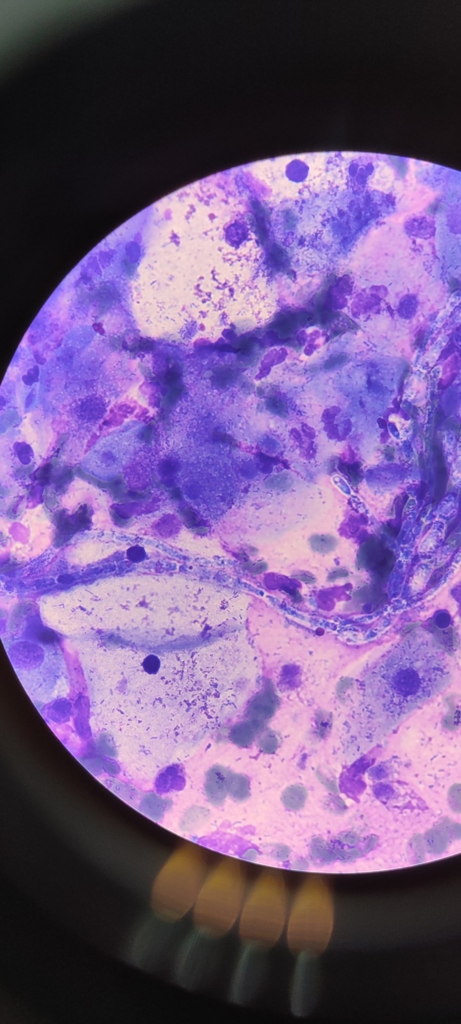
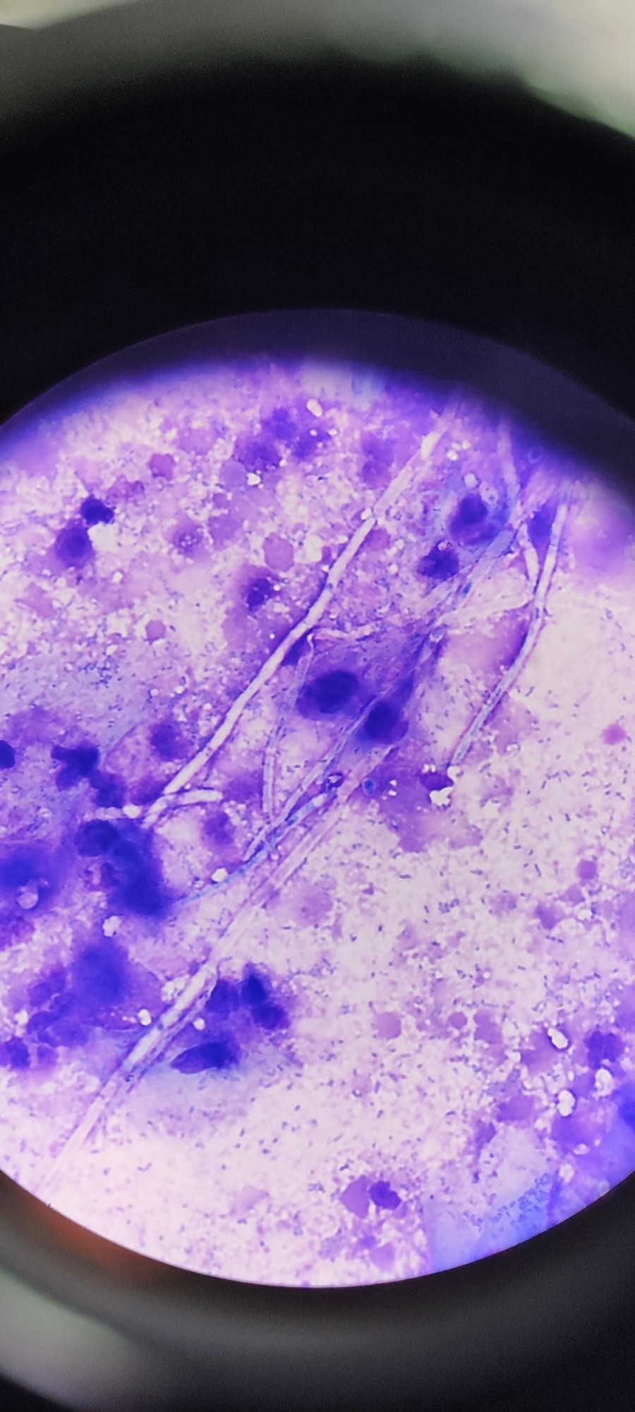
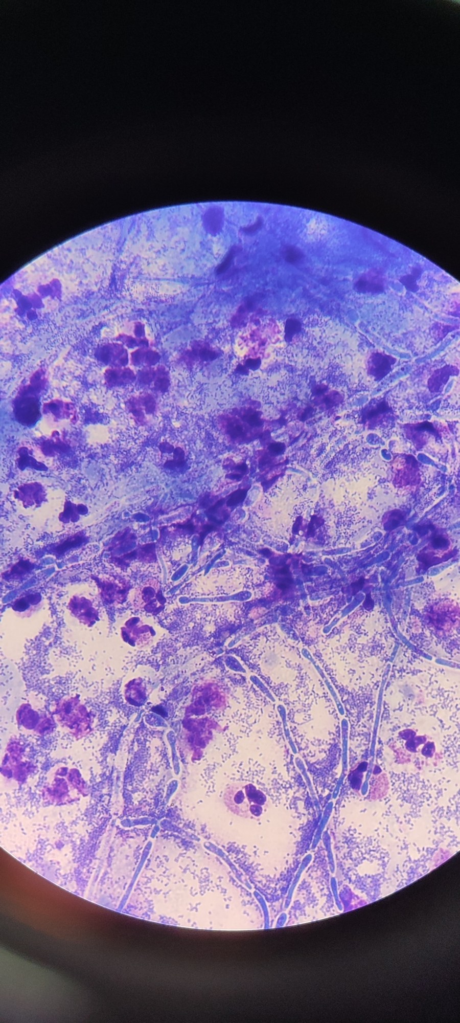
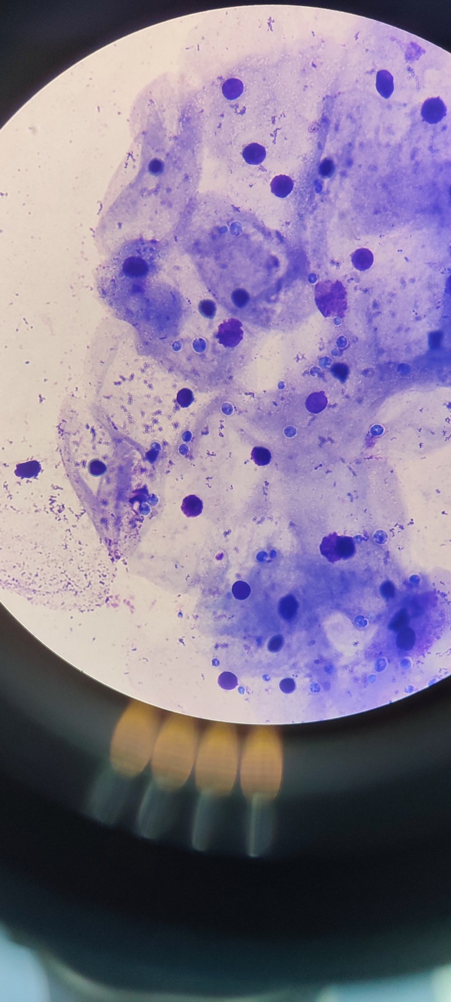
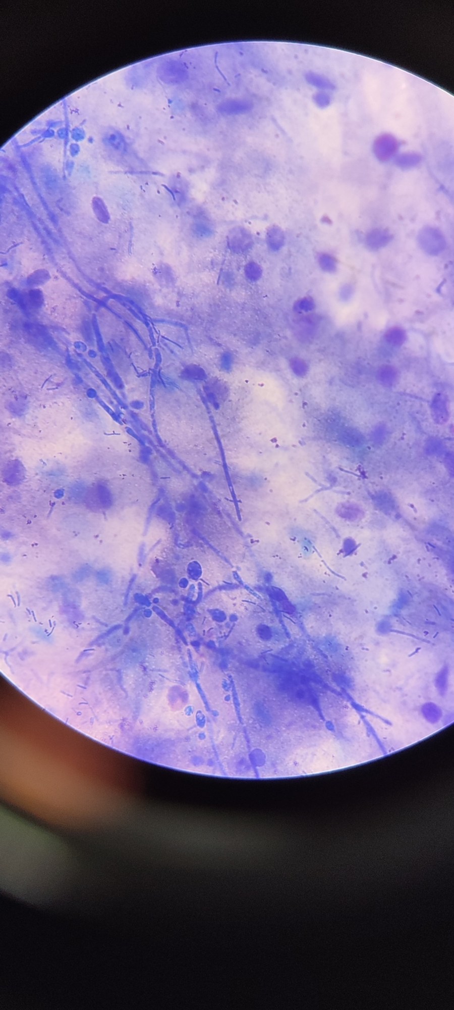
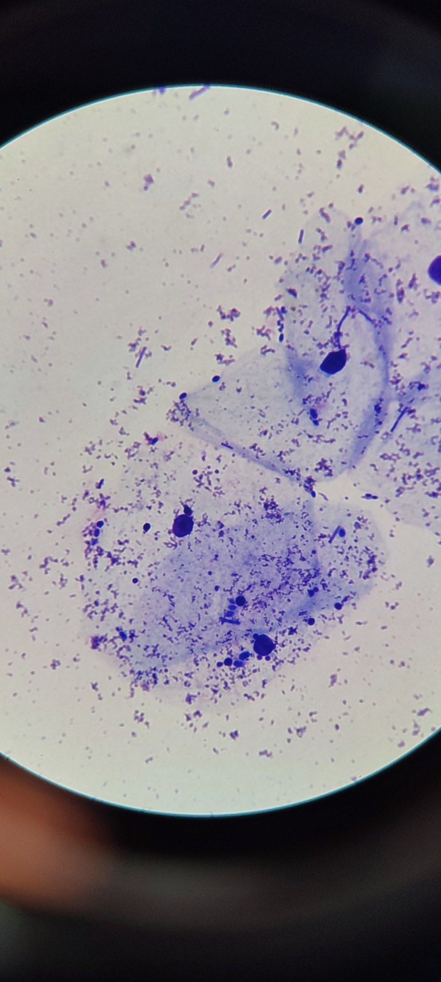
Reference information: 🔬 Yeast-like fungi detected during microscopy of a vaginal smear.
Morphology of Candida yeast-like fungi in a vaginal smear: pseudomycelium, blastospores, and co-occurring microflora during microscopy of a flora smear.
Autumn, cold, rain, “fungi”.
🙏 Images kindly provided by Clinical Laboratory Diagnostics Physician Polina Dybka, Krasnodar.
Reference information: 🔬 Yeast-like fungi detected during microscopy of a vaginal smear.

Microscopy of a vaginal smear in candidiasis: detection of pseudomycelium and blastospores of Candida fungi in a pregnant patient.

Microscopy of genital discharge: detection of pseudomycelium and Candida spp. yeast cells using methylene blue stain. A clinical case.

Analyzing the cytological picture of a male urethral smear during gonorrhea treatment control: morphology of columnar epithelium and leukocytes.