

🔬 Urine sediment microscopy. Now with captions by popular request! (View without captions→)
👤 Patient: 63 years old 🏥 Endocrinology Department ⚠ Protein in urine: 3 g/L
🙏 Images and description kindly provided by biologist Anna Feditskaya, Lugansk
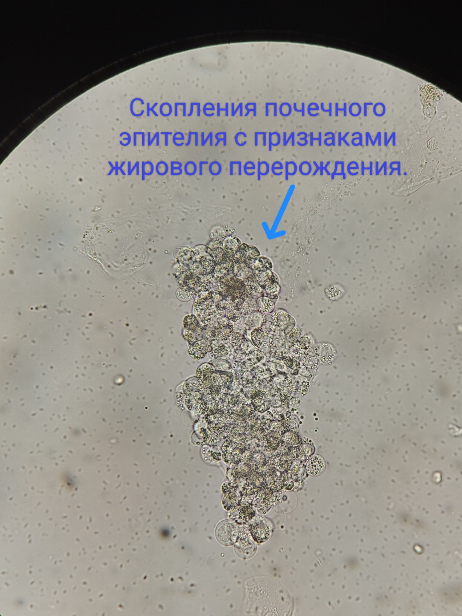
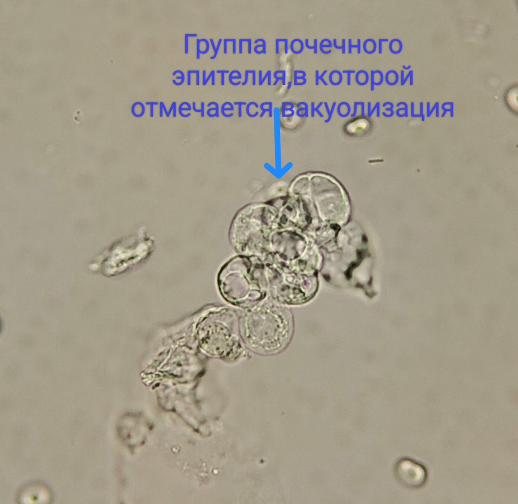
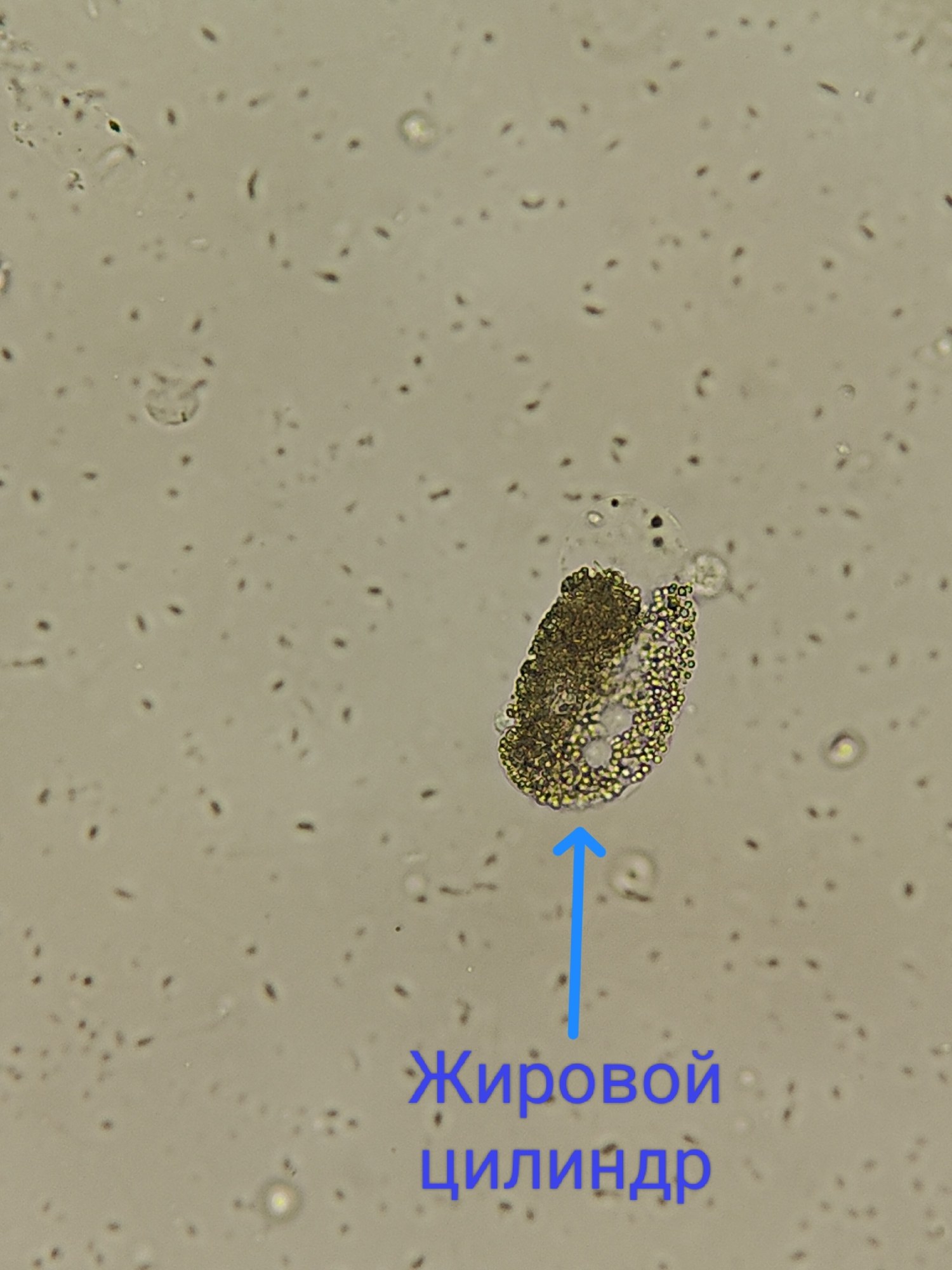
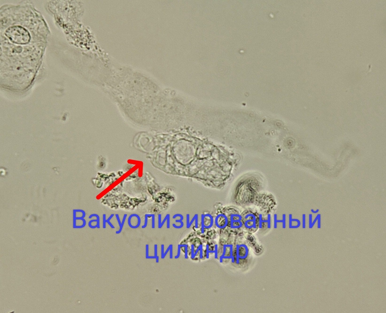
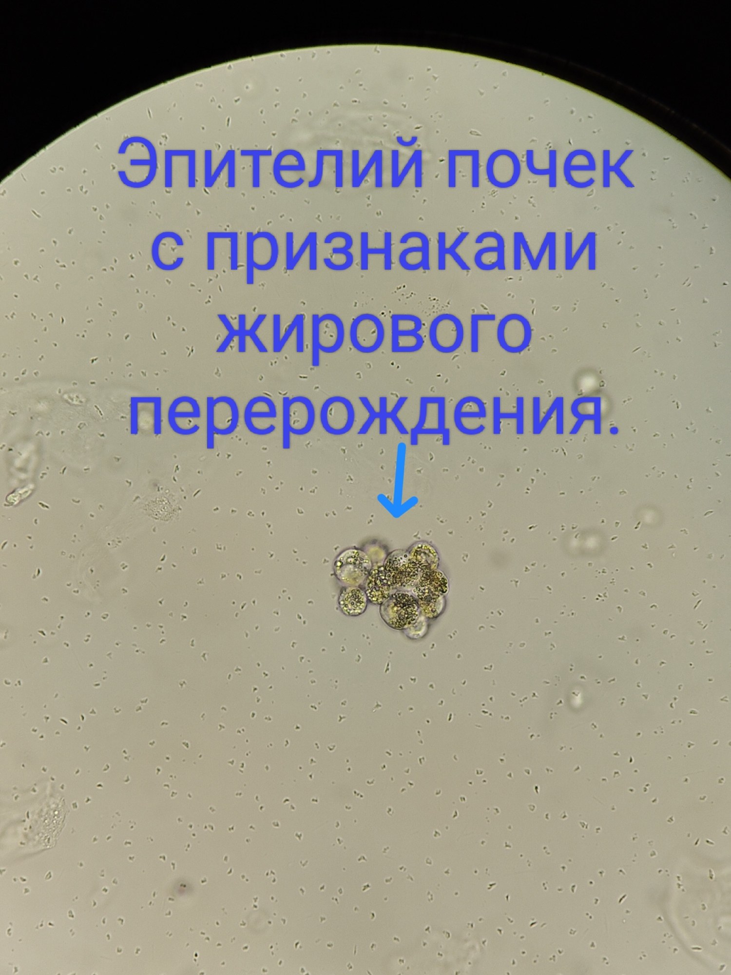
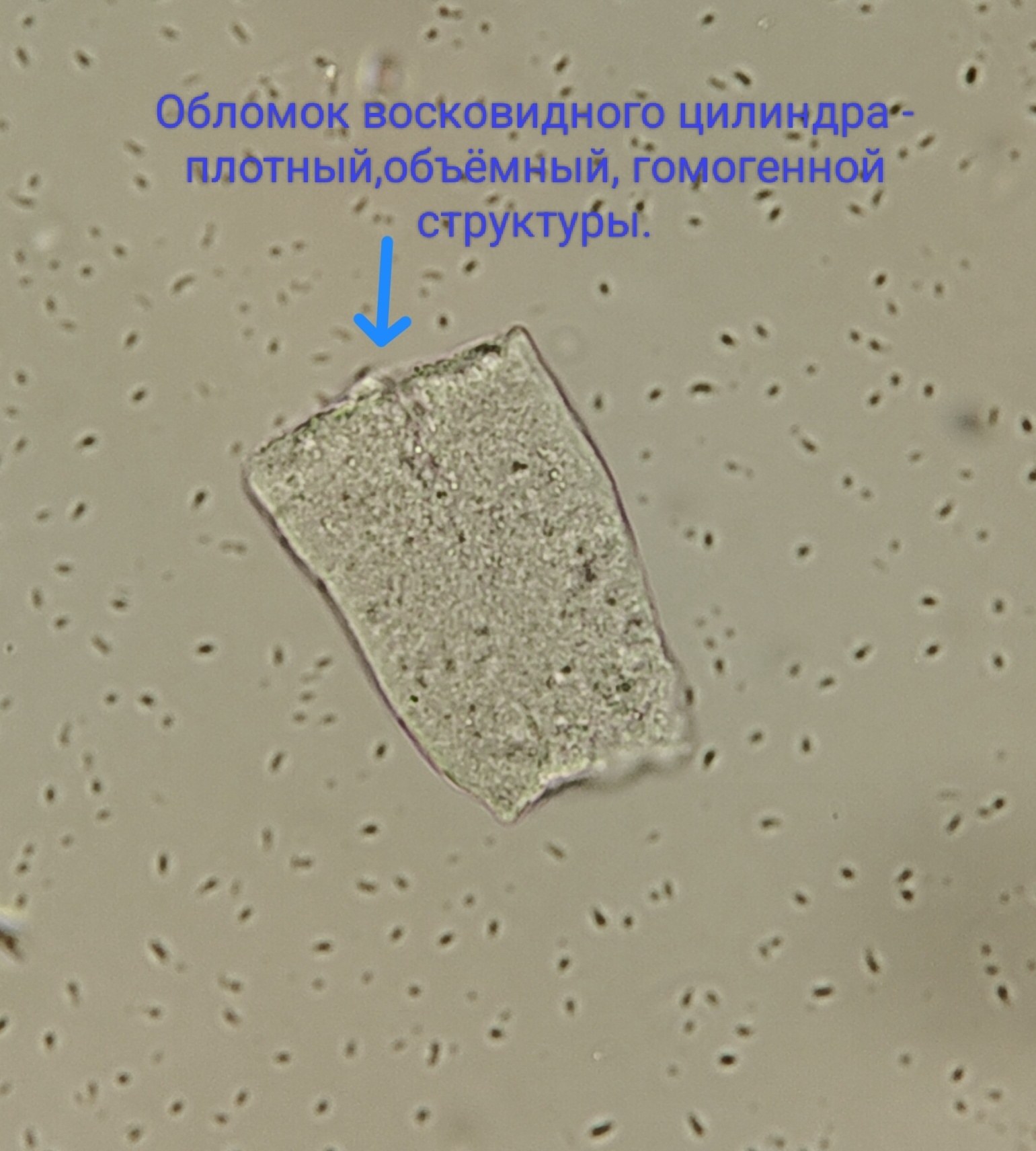
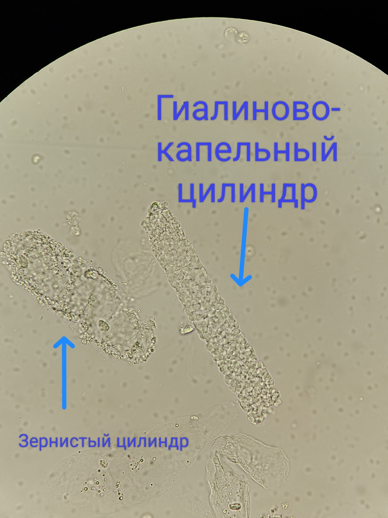
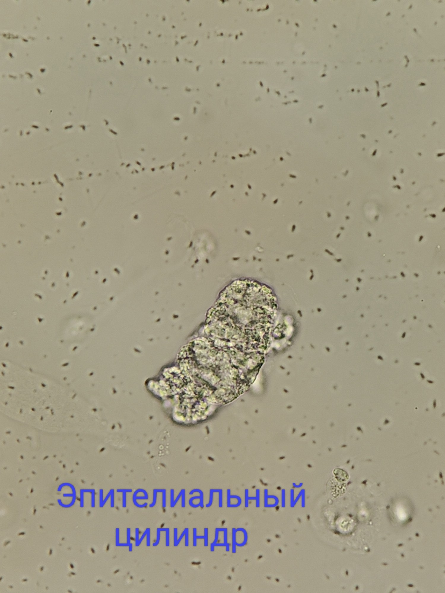
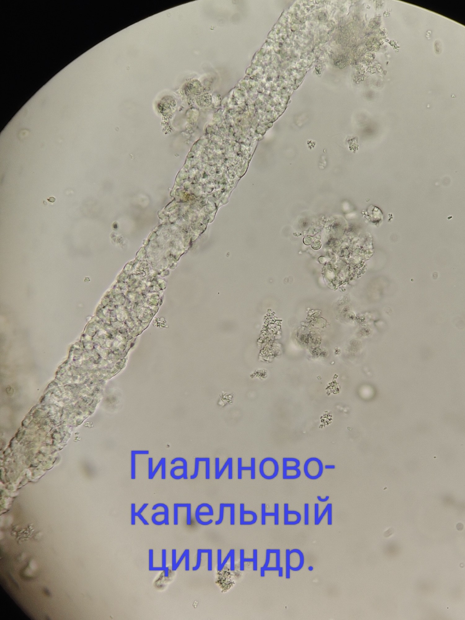
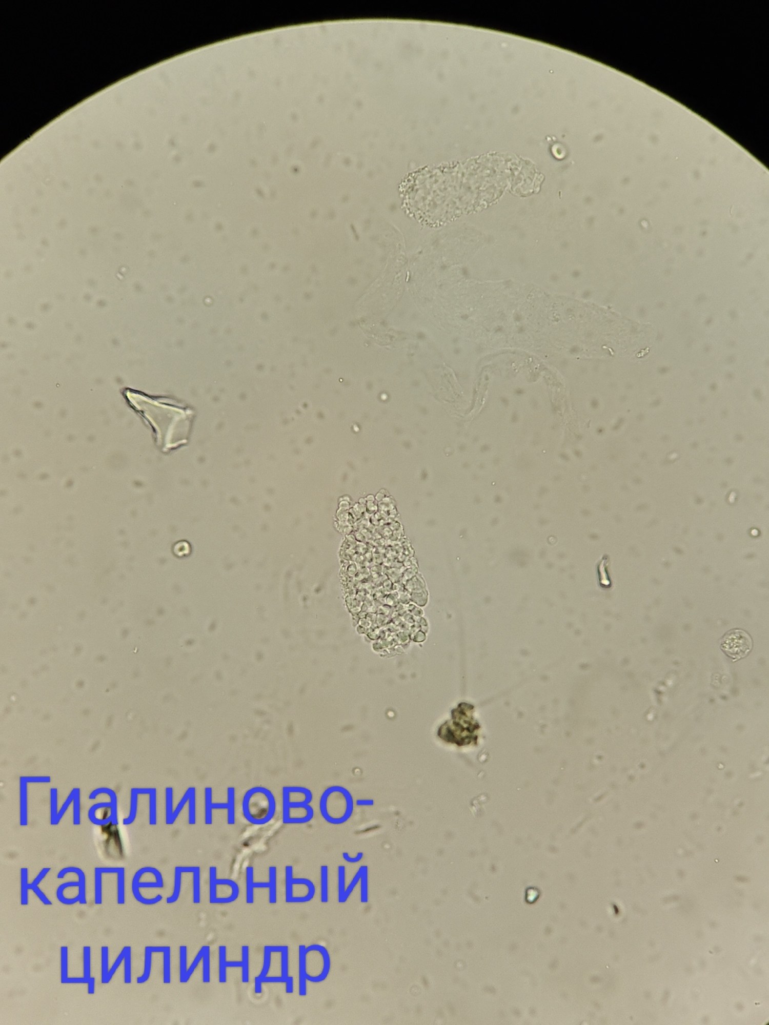
Atlas of urine sediment in proteinuria (3 g/L): microscopy of fatty, waxy, granular, and epithelial casts, as well as renal epithelial cells.
🔬 Urine sediment microscopy. Now with captions by popular request! (View without captions→)
👤 Patient: 63 years old 🏥 Endocrinology Department ⚠ Protein in urine: 3 g/L
🙏 Images and description kindly provided by biologist Anna Feditskaya, Lugansk

Urine sediment microscopy in a patient with proteinuria (3 g/L): fatty, granular, and epithelial casts, vacuolization and fatty degeneration of renal epithelium.

Urine sediment microscopy in a nephrology patient: differential diagnosis of casts, proteinuria 3 g/L, hematuria, and renal epithelial cells.

Epithelial casts in urine: microscopic examination of sediment, morphology of renal epithelial cells, diagnostic significance in tubular necrosis and nephritis.