

🔬 Someone is hiding here! Did you find it? Urine sediment microscopy.
🙏 Photo kindly provided by Clinical Laboratory Diagnostics Physician, Anna Ivanova, Moscow
Peculiarities of urine sediment microscopy in pronounced erythrocyturia: detection of barely visible elements, hyaline casts, and epithelial cells.
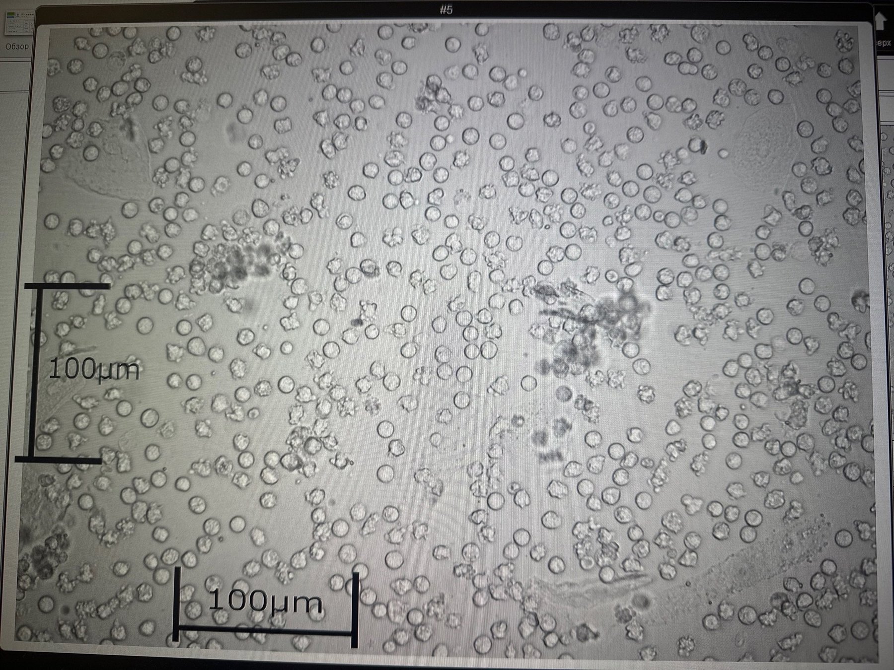
🔬 Someone is hiding here! Did you find it? Urine sediment microscopy.
🙏 Photo kindly provided by Clinical Laboratory Diagnostics Physician, Anna Ivanova, Moscow

Morphology of erythrocyte casts in urine sediment: microscopic signs of renal hematuria in glomerulonephritis and other kidney lesions.

Learn about the microscopic features of erythrocytes in stool. Understand why cells deform, leach, or retain shape in intestinal and gastric bleeding.

Microscopy of urine sediment: detection of spores and budding cells of yeast-like fungi (Candida spp.) in laboratory diagnostics of urogenital candidiasis.