

🔬Skin scales. Mycosis. Microscopy x200
🙏Material kindly provided by KDL biologist Tatiana Ivashchenko
Microscopic examination of skin scales in mycosis: identifying mycelial threads and fungal spores in a native preparation at 200x magnification.
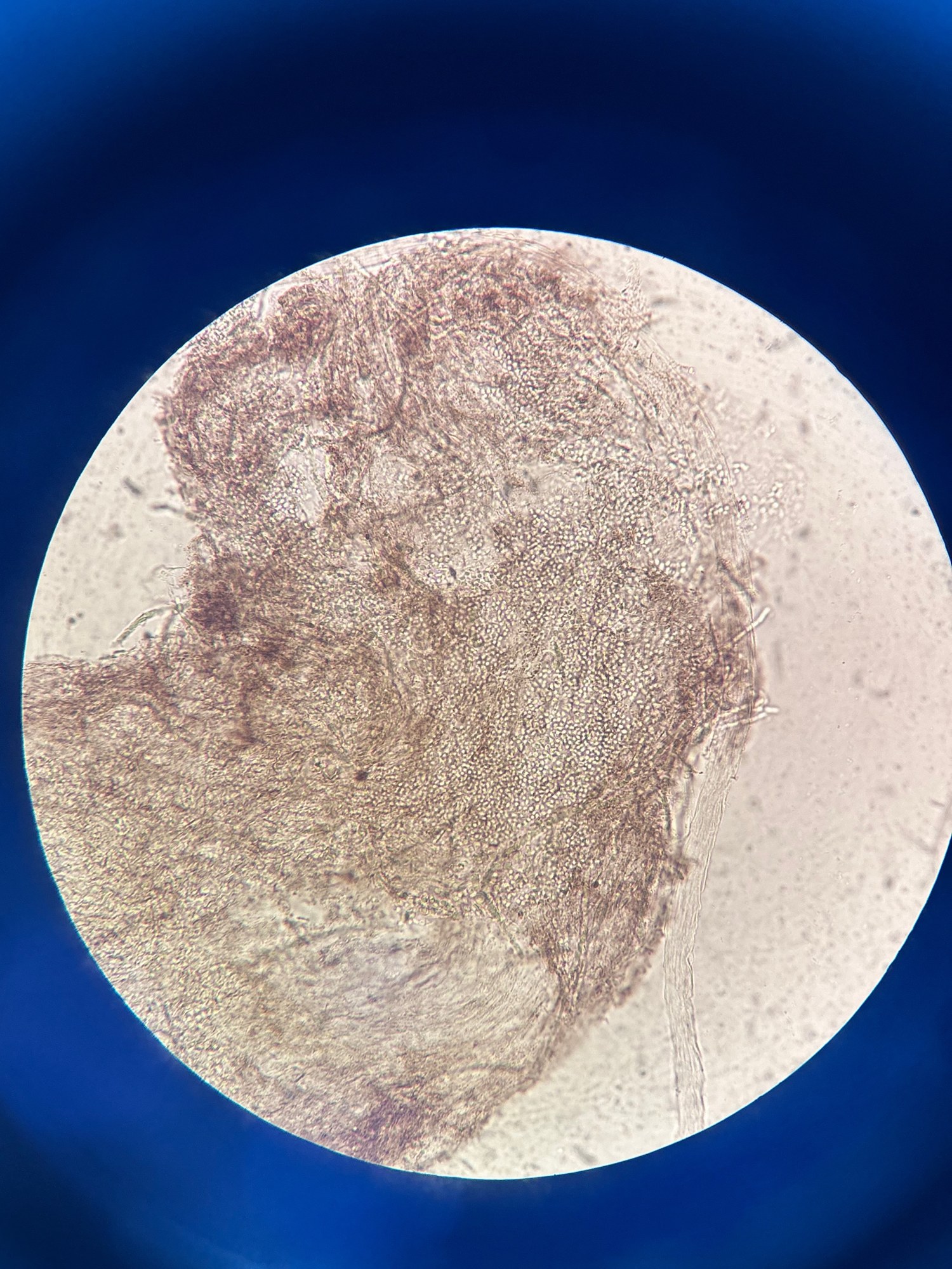
🔬Skin scales. Mycosis. Microscopy x200
🙏Material kindly provided by KDL biologist Tatiana Ivashchenko

Examples of skin and nail scraping microscopy in mycoses: visualization of mycelial threads, arthrospore chains, and pathogenic fungal elements in native preparations.

Microscopic examination of a nail plate scraping for suspected onychomycosis. Detection of branching fungal mycelial threads in a native preparation.

Microscopic view of a skin scraping from the groin folds: clusters of fungal mycelium filaments in a native preparation, indicative of inguinal dermatophytosis (epidermophytosis).