
🔬 Trichomonads with “legs” 🐾
🙏 Photos kindly provided by: Ispanka Isaeva
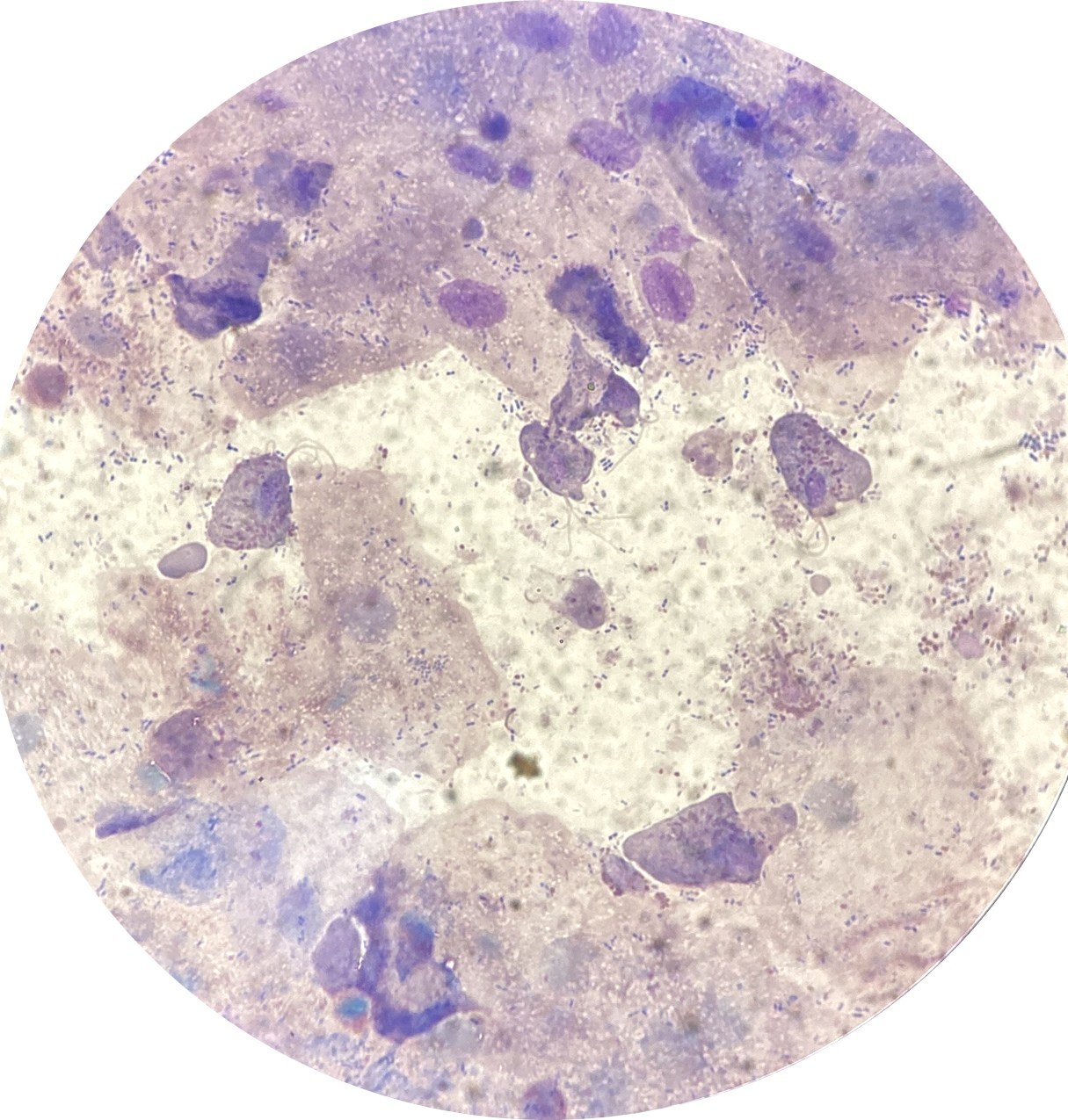
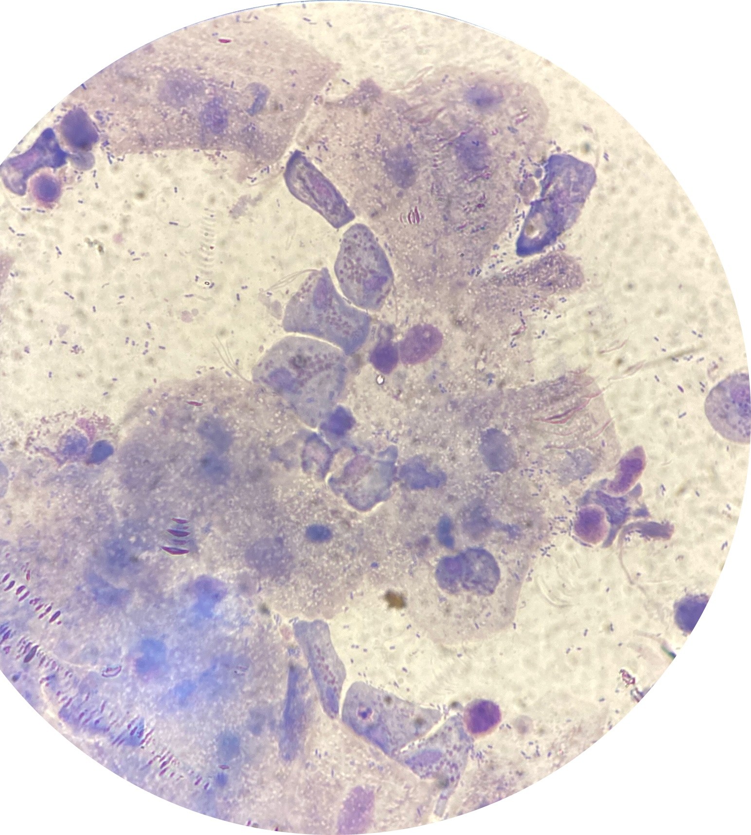
Key features of Trichomonas vaginalis microscopy in stained urogenital smears: identifying trophozoites, flagella, and coexisting bacterial flora.
🔬 Trichomonads with “legs” 🐾
🙏 Photos kindly provided by: Ispanka Isaeva

Microscopic view of a urogenital smear: identifying Trichomonas vaginalis trophozoites among squamous epithelial cells and abundant microflora.

Microscopic appearance of Trichomonas vaginalis in a stained male flora smear: leukocyte reaction, epithelial cells, and PCR confirmation.

Morphological features of Trichomonas vaginalis in a flora smear stained with Romanowsky stain: detection of trophozoites amidst leukocyte infiltration.