

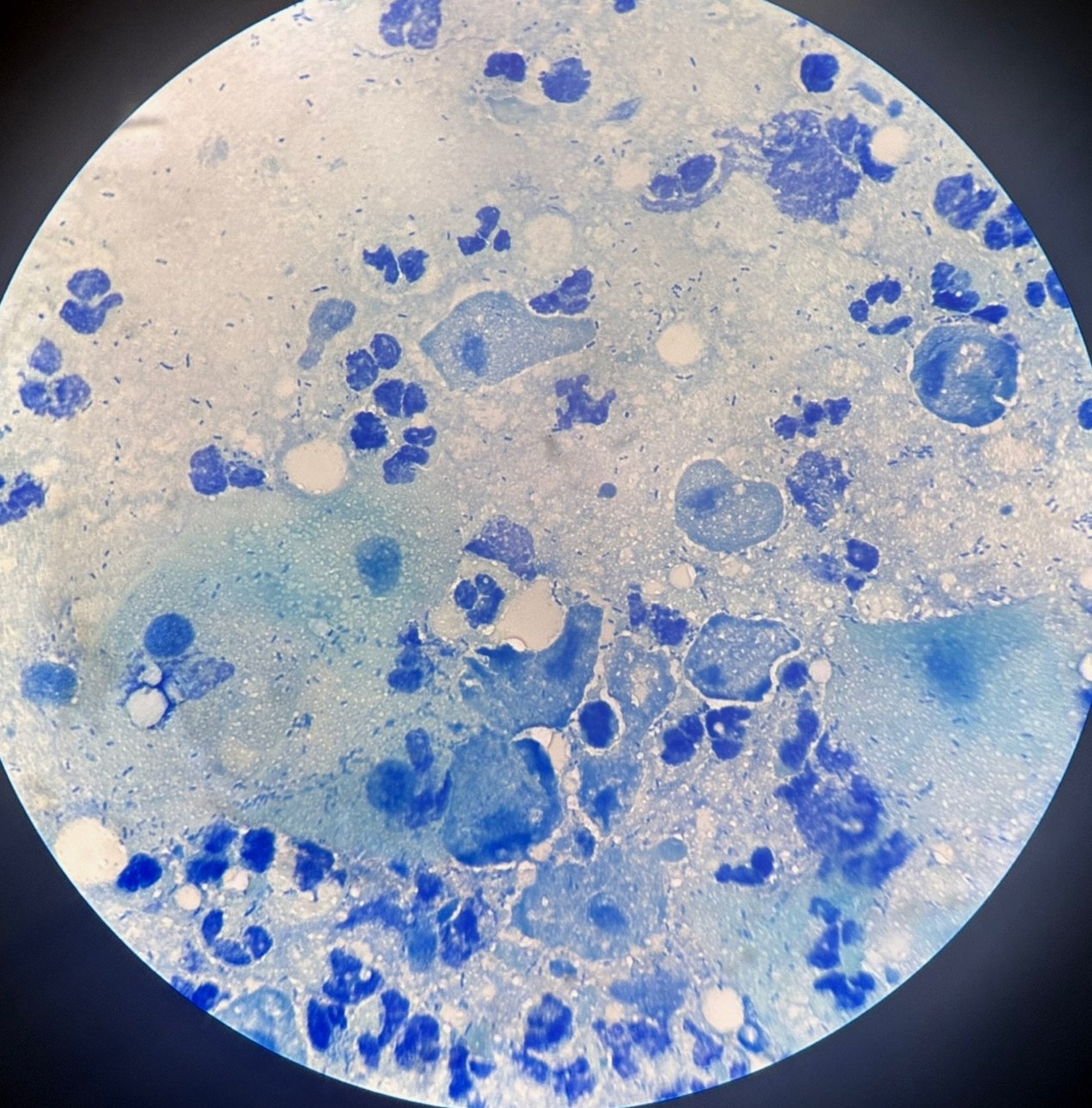
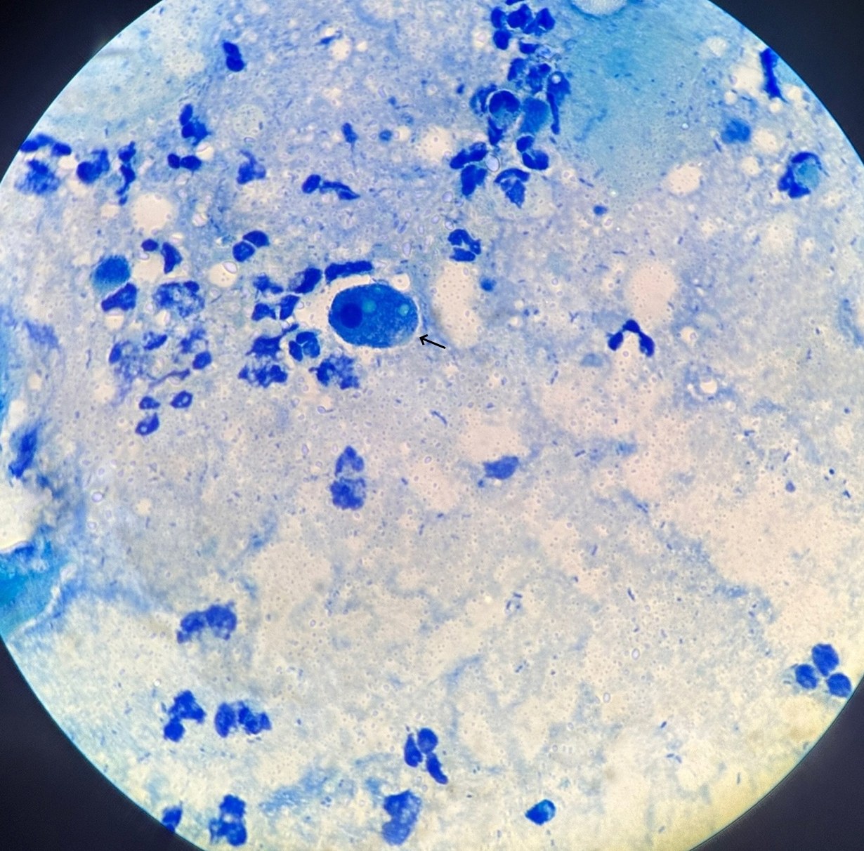
🔬 Microscopy of a vaginal smear from a 21-year-old girl. Trichomonas vaginalis. Colleagues, what do you think the object indicated by the arrow in the 2nd image is?
🙏 The images were kindly provided by clinical laboratory diagnostics physician, Anna Ivanova Moscow


