

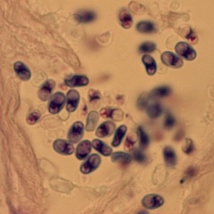
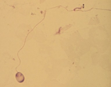
Microsporidia spores from a corneal section, stained with Giemsa. 2nd image in the carousel: A single microsporidia spore from the same case as in the first image, stained with Giemsa. In this image, the polar tube is extruded.
There are at least 15 species of microsporidia that have been identified as human pathogens; the vast majority of cases are caused by Enterocytozoon bieneusi, followed by some Encephalitozoon species (E. cuniculi, E. hellem, E. intestinalis (= Septata intestinalis)). Other less common causative agents include members of the genera Anncaliia (= Brachiola) (A. algerae, A. connori, A. vesicularum), Microsporidium (M. ceylonensis, M. africanum), Trachipleistophora (T. hominis, T. anthropophthera), Nosema ocularum, Pleistophora ronneafiei, Vittaforma corneae (= Nosema corneae), Tubulinosema acridophagus, and an unknown species, probably belonging to Endoreticulatus.
Microsporidia are a group of unicellular intracellular parasites closely related to fungi, although the nature of the relationship with the kingdom Fungi is not clear. The taxonomic position of this group has been repeatedly discussed and revised; historically, they were considered protozoa and often remain under the purview of diagnostic parasitology laboratories.
💎 Find more useful materials on parasitology in our LAB-TESTS library. The link with access to the library is automatically sent to you via private message after paying for a VK donut Labformula subscription.


