

Mitotic figures in the peripheral blood of cats diagnosed positive for FelV, FIV, 1000x magnification
🙏 Photos kindly provided by Amira Kuznetsova, hematologist at the VETLAB veterinary laboratory network, Moscow Amira Kuznetsova
Mitotic figures in the peripheral blood of cats with Feline Leukemia Virus (FeLV) and Feline Immunodeficiency Virus (FIV). Features of blood smear microscopy in veterinary medicine.
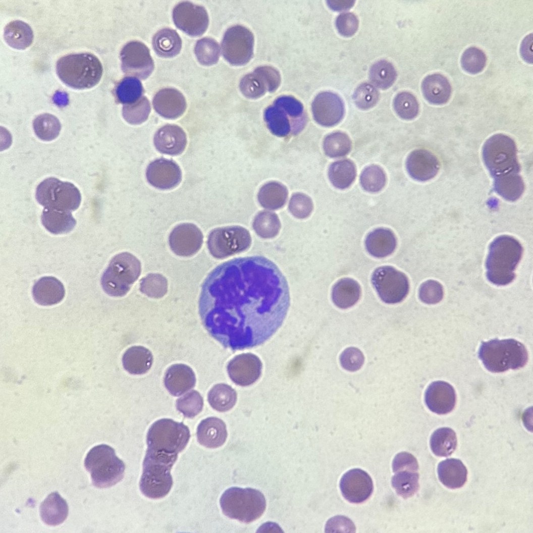
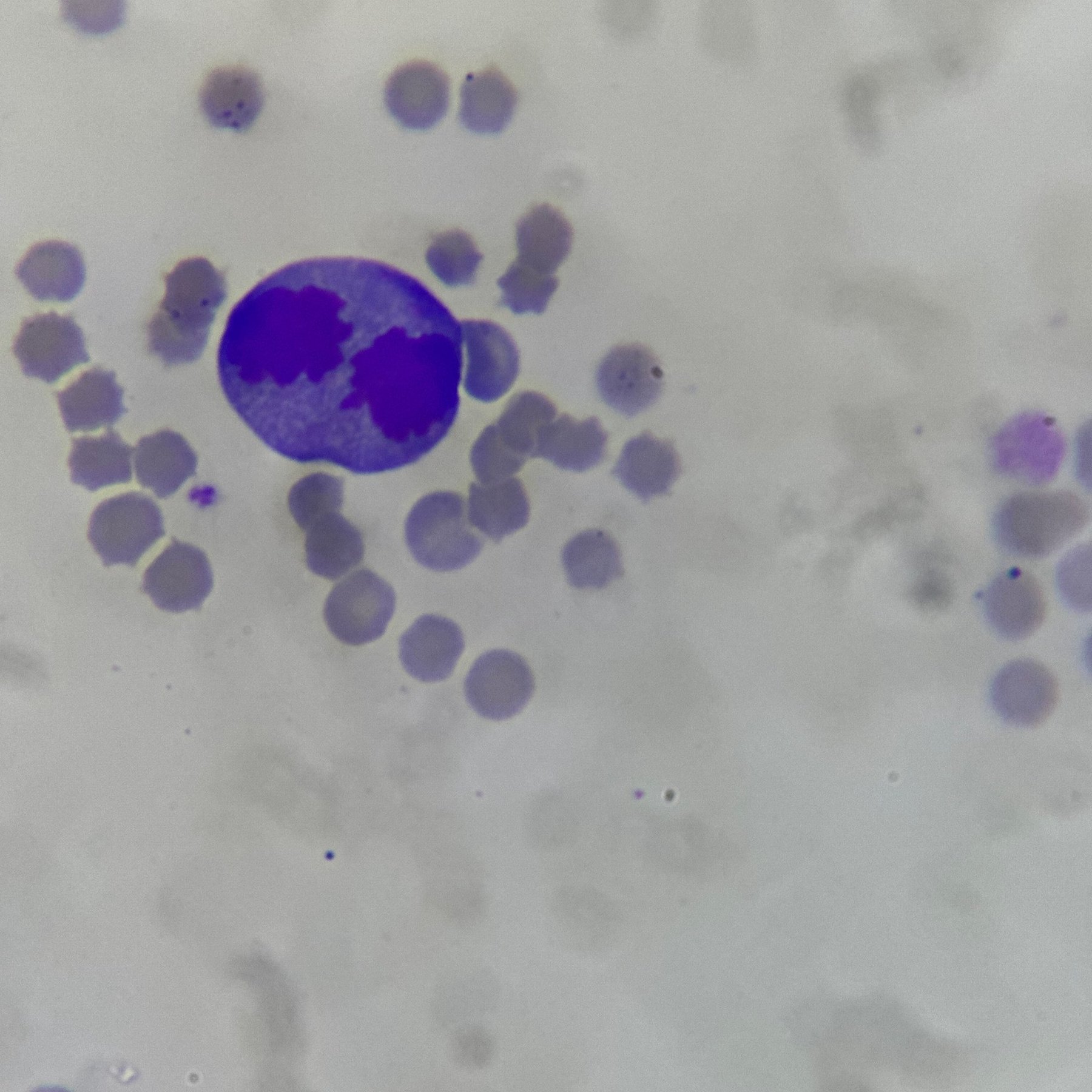
Mitotic figures in the peripheral blood of cats diagnosed positive for FelV, FIV, 1000x magnification
🙏 Photos kindly provided by Amira Kuznetsova, hematologist at the VETLAB veterinary laboratory network, Moscow Amira Kuznetsova

Morphology of a hypersegmented neutrophil in a blood smear from a 22-year-old cat. Features of nuclear hypersegmentation in geriatric animals in veterinary medicine.

Explore the morphological features of peripheral blood basophils in ferrets, dogs, and cats. Microscopy of blood smears stained with Romanowsky-Giemsa at 1000x magnification.

Explore the unique morphology and granularity of eosinophils in peripheral blood smears of mammals: a comparative microscopy guide for veterinary hematology.