

Urinary Amoebiasis: A Rare Manifestation of Entamoeba histolytica Infection
Urinary amoebiasis is a rare disease caused by the parasite Entamoeba histolytica. This parasite exists in two forms: the infectious cyst and the active form – the trophozoite. The disease can occur when the parasite enters the bladder via the bloodstream, lymph, or directly from the intestines. Clinical manifestations include hematuria, dysuria, suprapubic pain, and fever. Diagnosis is based on cytological and microscopic examination of urine, histological analysis, and serological confirmation. Treatment is carried out using metronidazole.
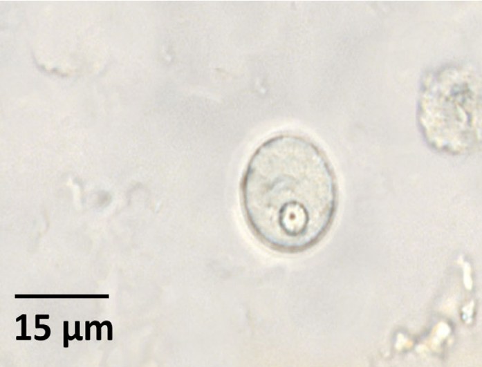
Figure 1: Urine sediment. The image shows an Entamoeba histolytica cyst. This is a rounded structure with a refractive wall, a visible nucleus inside, measuring about 15 µm. The image was taken at ×1000 magnification (wet mount).
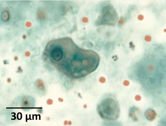
Figure 2: Cytological smear from urine. Entamoeba histolytica trophozoite, Papanicolaou stained. Greenish cytoplasm with irregular contours resembling pseudopods is visible, along with vacuoles containing phagocytosed erythrocytes. A nucleus with a prominent nucleolus is also clearly visible. Magnification ×400.
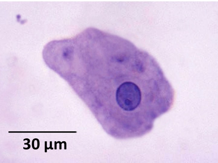
Figure 3: Cytological smear from urine. Entamoeba histolytica trophozoite, stained with Wheatley’s trichrome method. The image demonstrates the typical morphology of the parasite. Magnification ×1200.
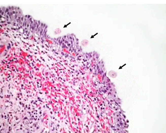
Figure 4: Histological section of the bladder. The mucous membrane is inflamed, with pronounced vascular hyperemia, epithelial loss, and ulcerative areas. In these areas, amoebae are visible near the epithelium (indicated by black arrows). Stained with hematoxylin and eosin, magnification ×100.
💎 Find more useful materials on parasitology in our LAB-TESTS library. The link with access to the library is automatically sent to you via private message after paying for a VK donut Labformula subscription.
Case source: Martínez‐Girón R., Martínez‐Torre S., Martínez‐Torre C. Urinary amoebiasis //Diagnostic Cytopathology. – 2020. – Т. 48. – №. 12. – С. 1313-1315.


