

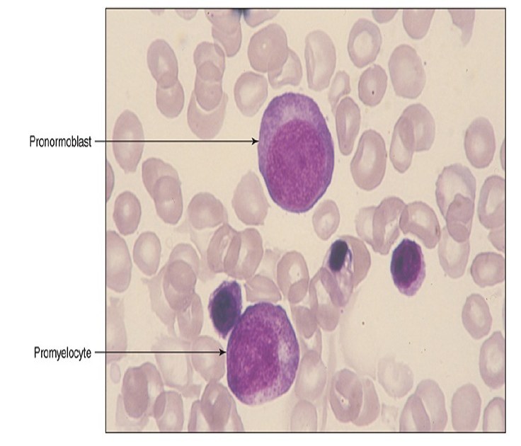
Pronormoblast vs. Promyelocyte
PRONORMOBLAST The nucleus is large, dark purple, and centrally located. Nuclear chromatin has fine stippling. The cytoplasm is intensely basophilic, with a light perinuclear area.
PROMYELOCYTE The promyelocyte nucleus is eccentrically located. Nuclear chromatin is finely condensed. The cytoplasm has numerous dark granules, which may obscure the nuclear contours.
Pronormoblast Nucleus is large, deep purple, and centrally located. Nuclear chromatin is finely stippled Cytoplasm is intensely basophilic light areas of mitochondria Light area next to the nucleus is the Golgi.
Promyelocyte Nucleus is purple and eccentrically located. Nuclear chromatin is finely condensed Cytoplasm has numerous dark, primary granules that have begun to obscure the nucleus.
Source: Andersons - Atlas of Hematology 2021 - 3 edition


