

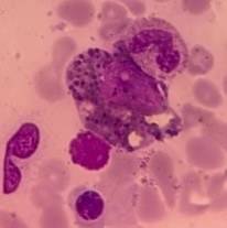
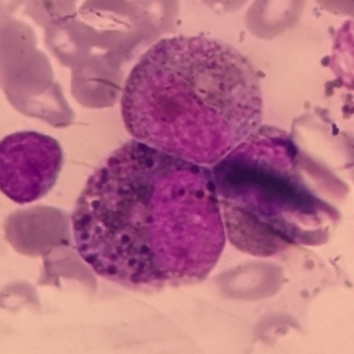
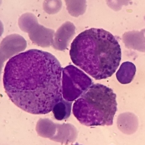
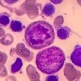
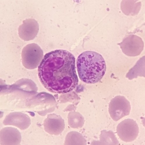
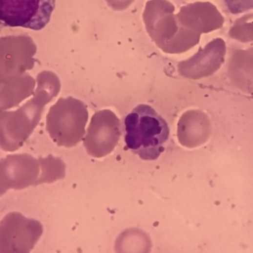
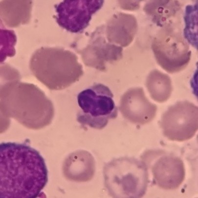
Archival slide from 2009. Bone marrow of a boy, 1 year 1 month old. Noteworthy features: 1-3 photos - Harlequin cells; 4 photo - mitosis of a myeloid cell; 5 photo - large basophilic stippling of an erythrocyte; 6-7 photos - abnormal, lobulated nucleus of an oxyphilic normoblast.
The child was diagnosed with MDS.
Romanowsky-Giemsa staining, x1000 magnification + phone camera magnification.
The material was kindly provided by clinical laboratory diagnostics doctor Nadezhda Petrenko, Blagoveshchensk.


