

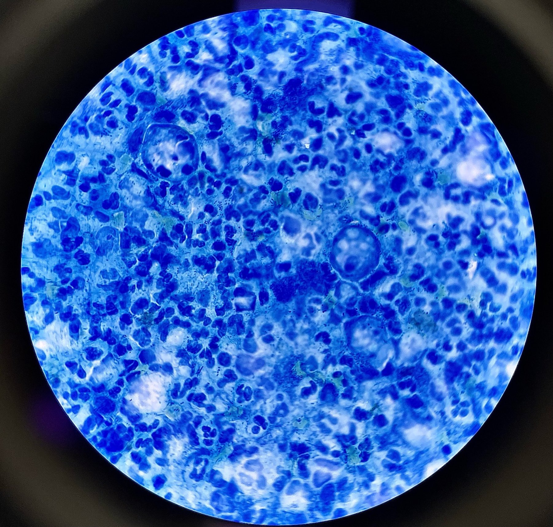
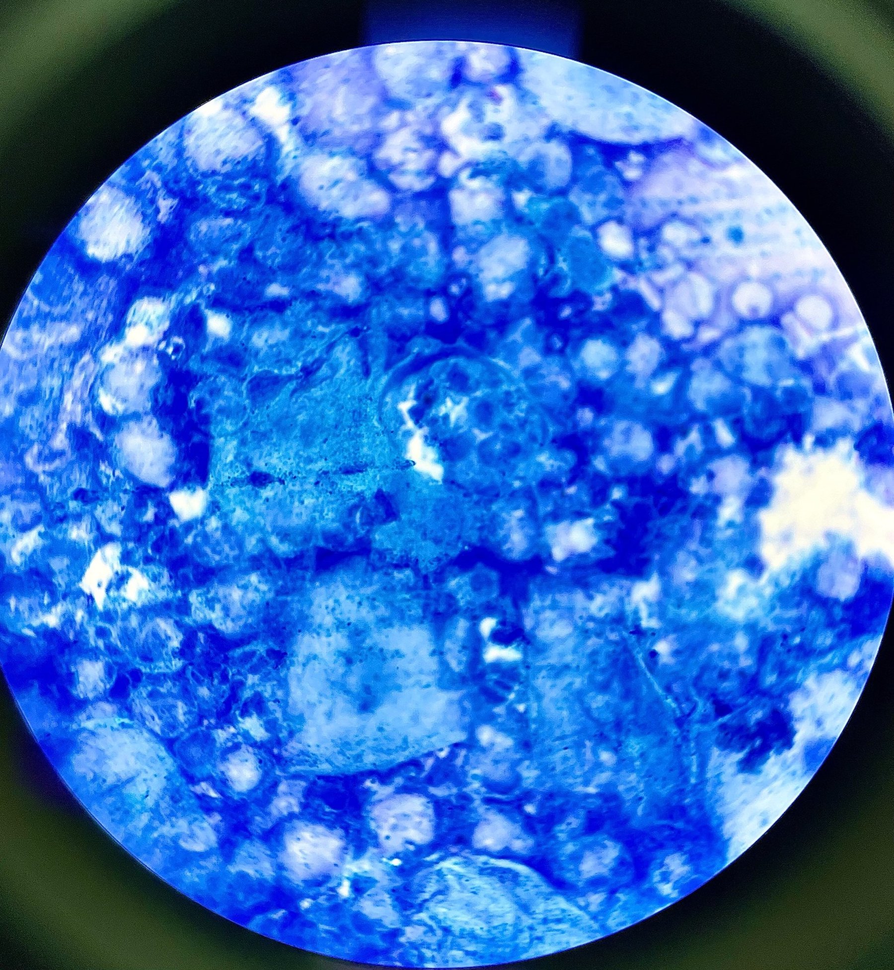
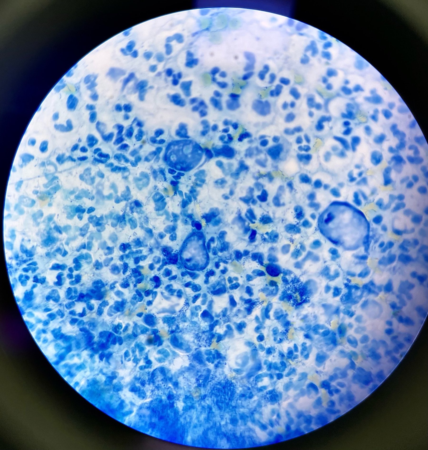
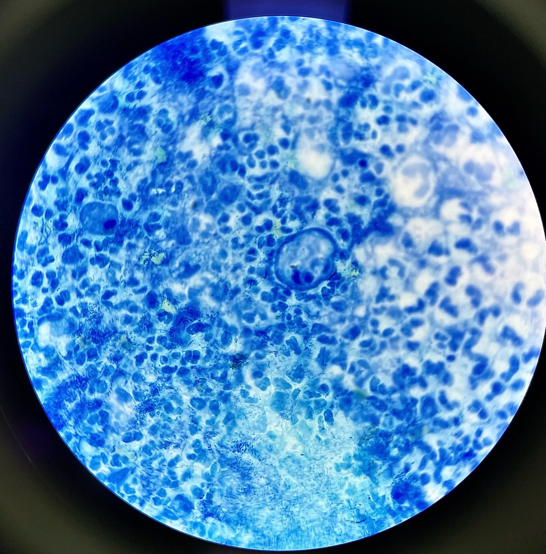
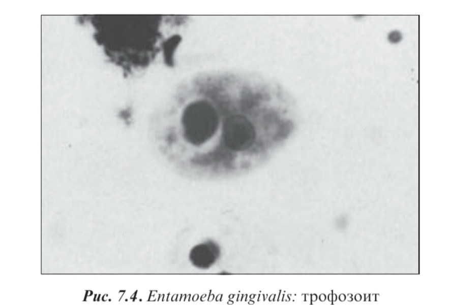
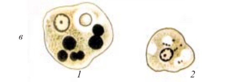
Oral amoeba (Entamoeba gingivalis). The trophozoite measures 6-30 µm; the cytoplasm is clearly divided into two layers. Phagocytosed bacteria and leukocytes at various stages of digestion can be seen within it. Localization: soft dental plaque, gingival pockets. The preparation was made from gum discharge. Staining with methylene blue, x1000 magnification
🙏 Material kindly provided for publication by - Clinical Laboratory Diagnostics Physician Elizaveta Emelyanova, Rostov-on-Don


