

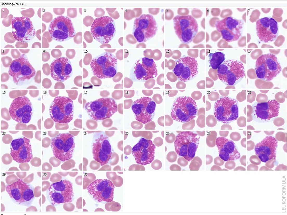
🔬 Eosinophils in blood smears.
👉 Eosinophils contain large orange granules in their cytoplasm, which stain intensely with eosin. The nucleus of eosinophils most often consists of two segments, which makes them easy to distinguish from other granulocytes.
🩸 Find a large database of photos with descriptions in our Library of Blood and Bone Marrow Secrets. The library volume is over 80 gigabytes! 💎 A link with access to the library is automatically sent to you via private message after paying for a VK donut Leukformula subscription.


