

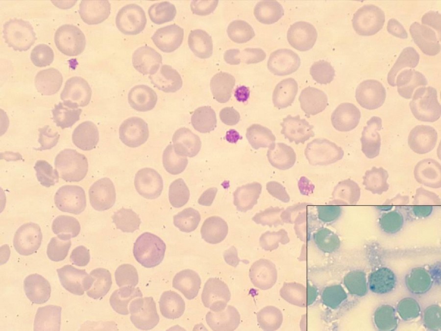
Poikilocytosis with the presence of acanthocytes, codocytes (target cells), and occasional eccentrocytes is observed. In the lower right corner (inset), an erythrocyte with typical precipitates, revealed by brilliant cresyl blue staining, is visible.
Clinical information: Peripheral blood smear of a 6-month-old girl of Pakistani origin diagnosed with Hemoglobin H disease (genotype -α3.7/–39.1).
Biological parameters: Hemoglobin: 8.2 g/dL MCV (mean corpuscular volume): 48.6 fL RDW (red cell distribution width): 26.9% No iron metabolism disorders detected.
Sample type: Peripheral blood
Staining type: May-Grünwald – Giemsa
Authors: Lourdes Castro Reyes, Marta Hidalgo Soto, Ana Ortuño Cabrero, David Benitez Pastor
Workplace: Hospital Vall D’Hebron


