

🔬Working in the Mediterranean region, I encounter thalassemias and pathological hemoglobins in practice, which I only saw in atlases in Russia. I’ll share my personal collection.
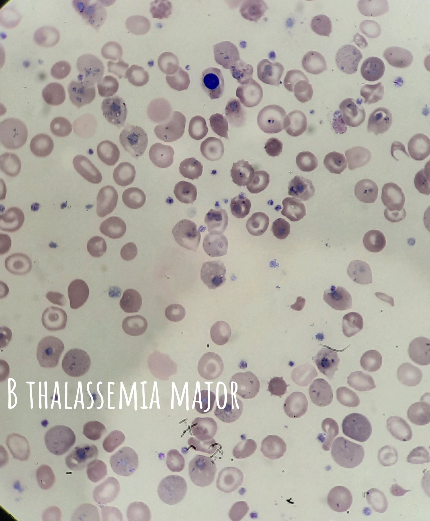
- B-thalassemia major Severe changes in the erythroid lineage: Anisocytosis and aniso-chromia of erythrocytes, Target cells, Normoblasts, Basophilic stippling, Pappenheimer bodies, Howell-Jolly bodies
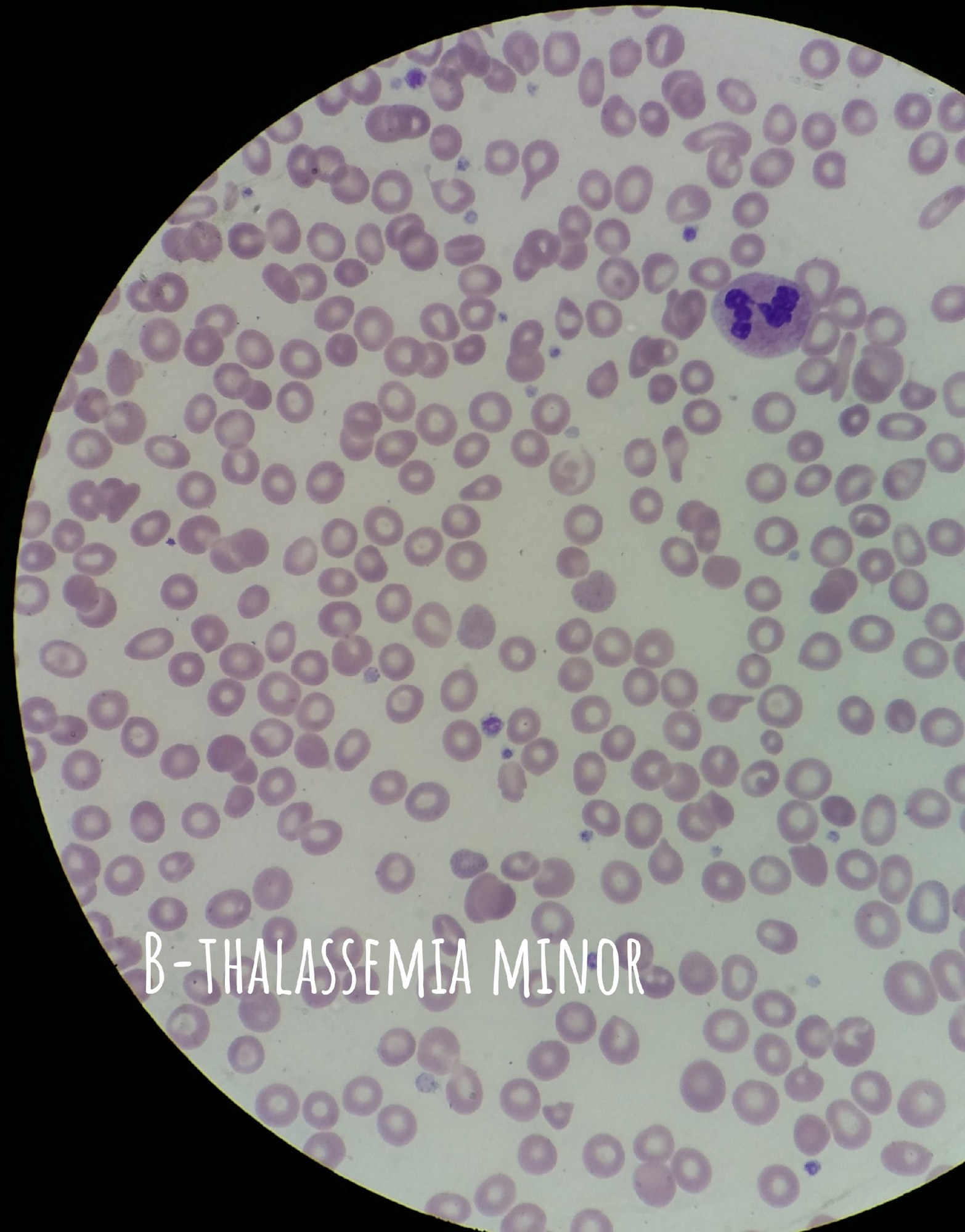
- B-thalassemia minor Very minor erythrocyte changes: Hypochromia and occasional target cells.
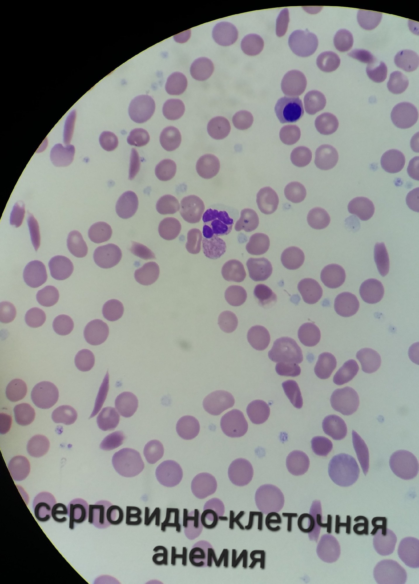
- Sickle cell anemia (Hemoglobinopathy S) Sickle cells
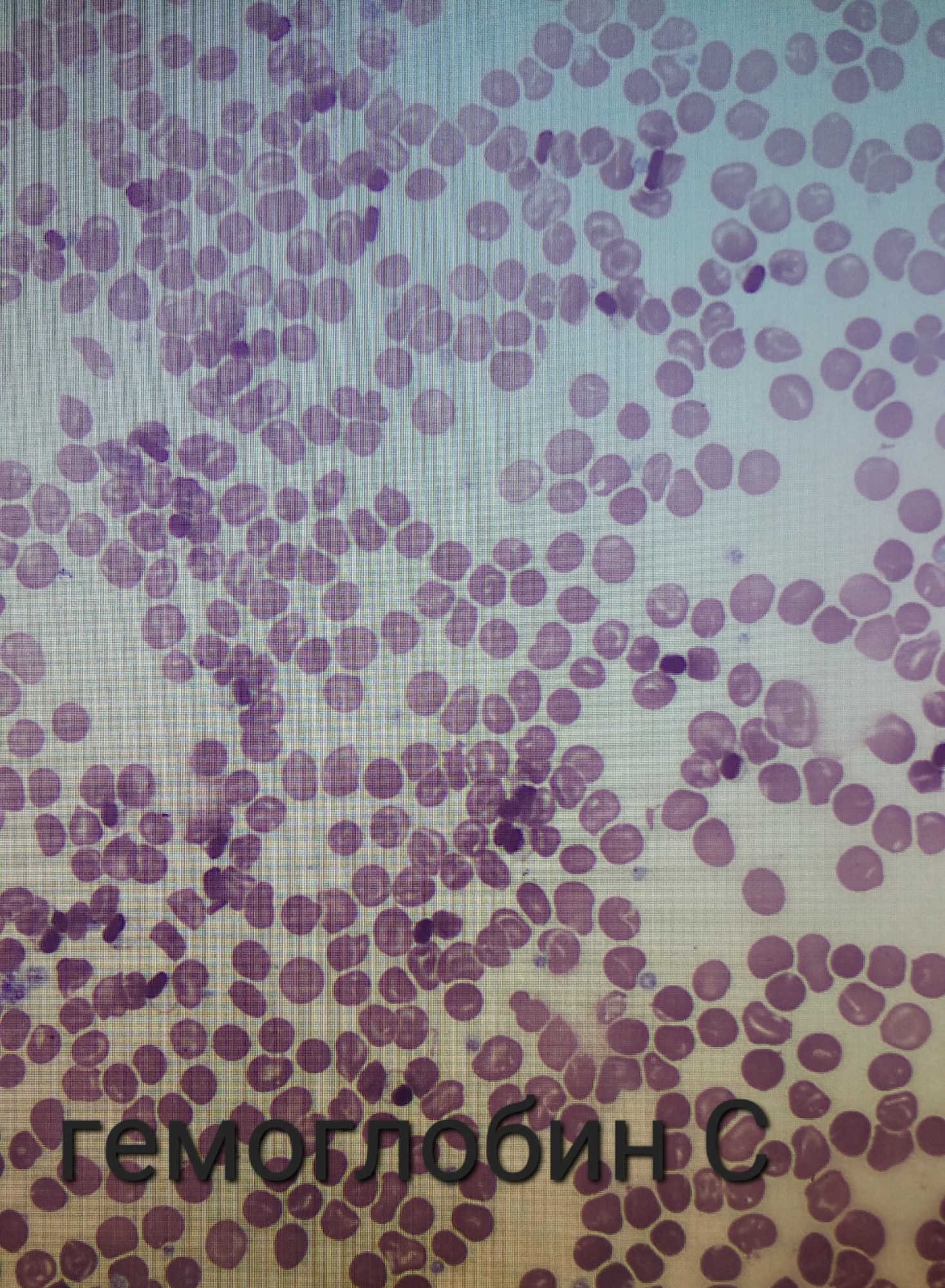
- Hemoglobinopathy C Characteristic crystals of pathological hemoglobin C in erythrocytes.
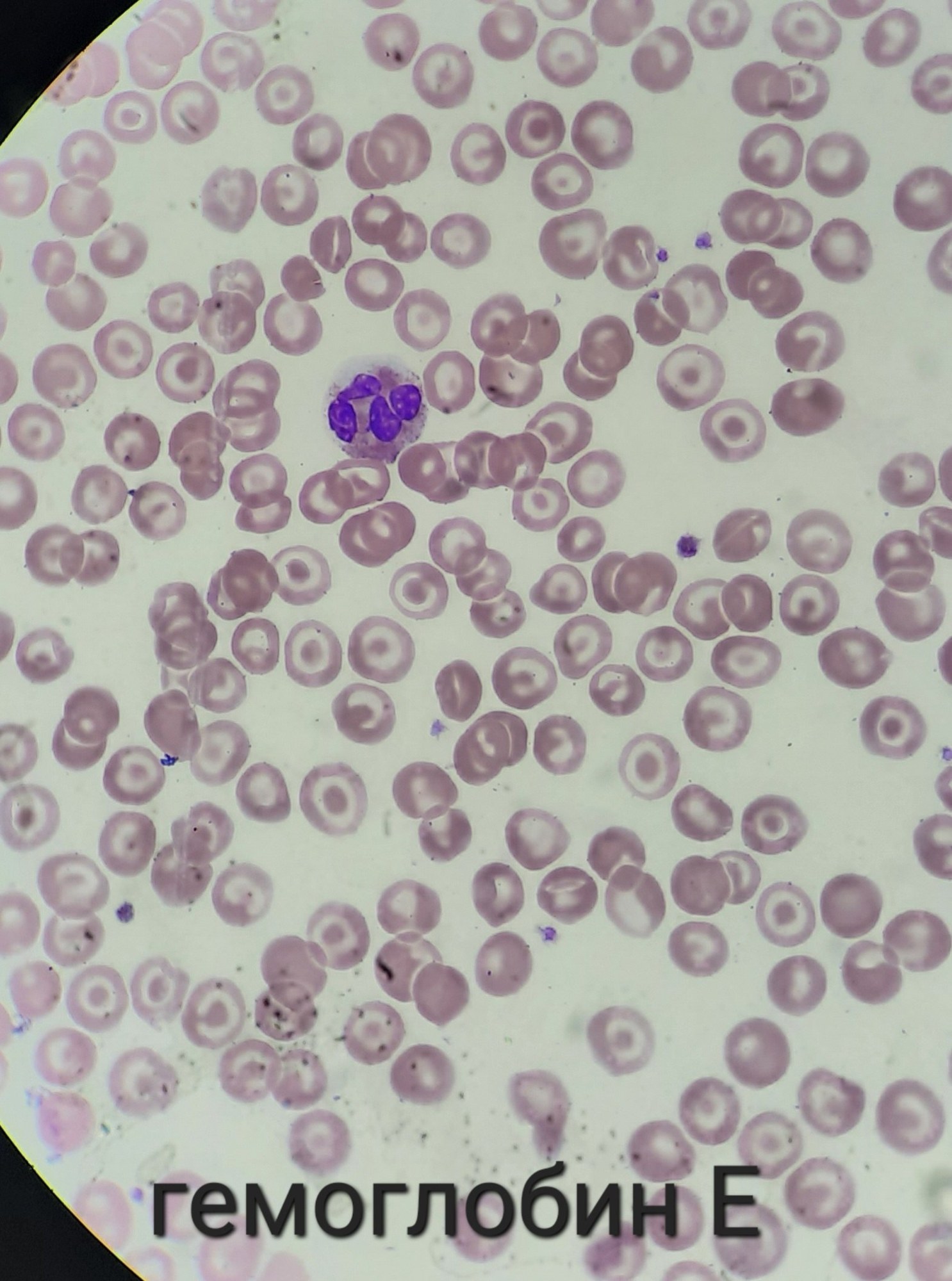
- Hemoglobinopathy E A migrant worker from Thailand. Almost all erythrocytes are target cells.
❓Does anyone know why there is no need to present a blood picture of Alpha-thalassemia?


