

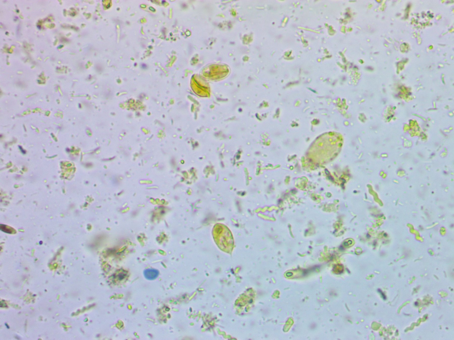
🔬 Three different parasites in one preparation! Let’s study the morphology of protozoa. ✅ The following structures were identified in the preparation:
- A cyst of Lamblia intestinalis (Giardia lamblia), and a trophozoite was found nearby, confirming the presence of the active form of the parasite.
- Below, a blastocyst of Blastocystis hominis was identified.
- And even further below, the presence of a trophozoite of Chilomastix mesnili was noted. Microscopic examination of a stool smear in Lugol’s solution (magnification ×600) 🙏The publication was kindly provided by Cand. Biol. Sci., Associate Professor, Parasitologist Vladimir Turitsyn


