

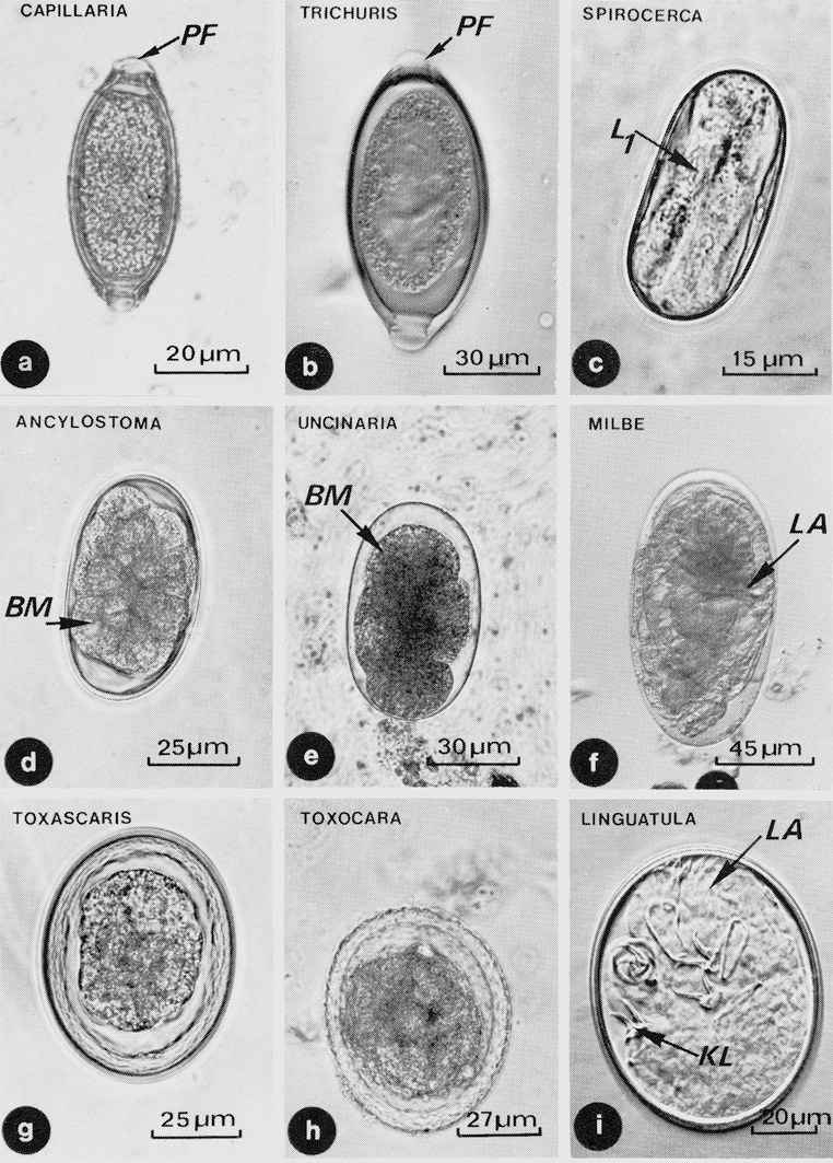
🔬Nematode eggs are found in dog feces. Note that Toxascaris leonina eggs (g) have a smooth surface, while Toxocara canis (h) have a rough surface. a – Capillaria putori, b – Trichuris vulpis, c – Spirocerca vulpis, d – Ancylostoma sp., e – Uncinaria sp., f – mite egg, g – Toxascaris leonina, h – Toxocara canis, i – Linguatula serrata, already containing a four-legged larva. Abbreviations: BM - Blastomeres, KL - Claws (talons) of legs, L1 - Nematode larva, LA - Linguatula larva, PF - Polar plug
Source: Mehlhorn H. - Encyclopedia of Parasitology, 4-ed, 2016


