

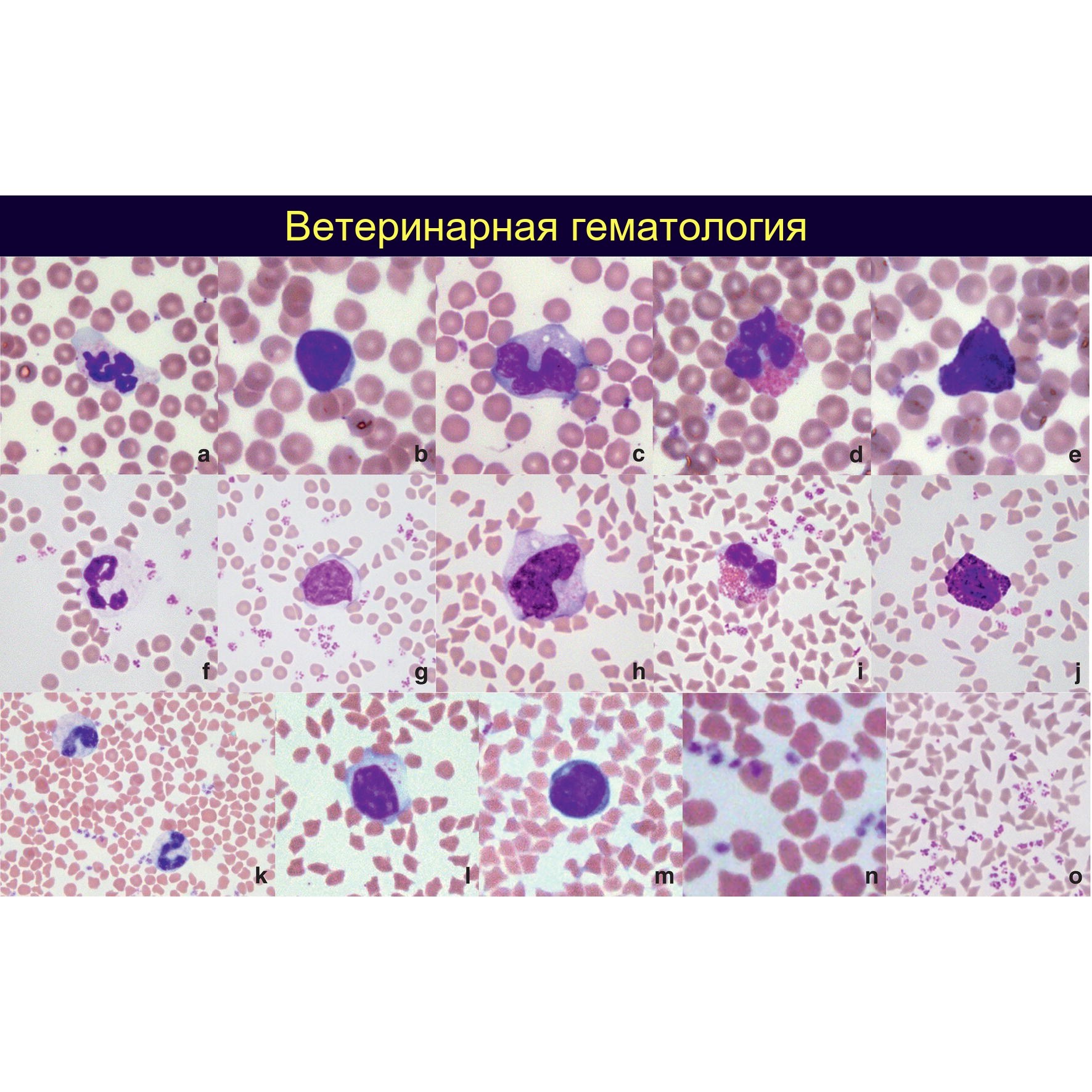
Comparative morphology of sheep and goat leukocytes and platelets (a) segmented neutrophil; (b) lymphocyte; (c) monocyte; (d) eosinophil; (e) basophil; (f) segmented neutrophil.
Goat leukocytes and platelets: (g) lymphocyte; (h) monocyte; (i) eosinophil; (j) basophil; (k) toxic band neutrophil with vacuolization and cytoplasmic basophilia; (l) granular lymphocyte.
Additional goat leukocytes: (m) lymphocyte with signs of reactivity: dark blue cytoplasm with a small pale perinuclear Golgi apparatus zone.
Wright–Giemsa stain.
(n) sheep platelets. (o) goat platelets.
Source: Brooks M. B. - Schalm’s Veterinary Hematology, 2022 🏛 Library “Veterinary Laboratory Medicine” ➡️ Level III subscription Level III - “Metamyelocyte” provides access to this book and the entire training course “Hematology of Exotic Animals”: Lesson 1: Reptile Blood Morphology 🦎 Lesson 2: Avian Blood Parasites 🐦 Lesson 3: Features of Blood in Domestic Exotic Pets🐷 Lesson 4: Differences Between Monkey and Human Blood 🐵
🔗 Details and subscription → vk.com/@leukoformula-3-level


