

Peripheral blood smear in myelofibrosis
Myelofibrosis, classified as “primary,” belongs to myeloproliferative neoplasms where fibrosis is reactive to a hematological neoplasm. Secondary myelofibrosis represents a bone marrow stromal reaction to a non-hematological disease, for example, it can occur in cases of carcinoma or sarcoma metastasis to the bone marrow. Both of these variants of myelofibrosis will have a rather similar picture upon examination of peripheral blood smears, with some exceptions, which we will consider in more detail.
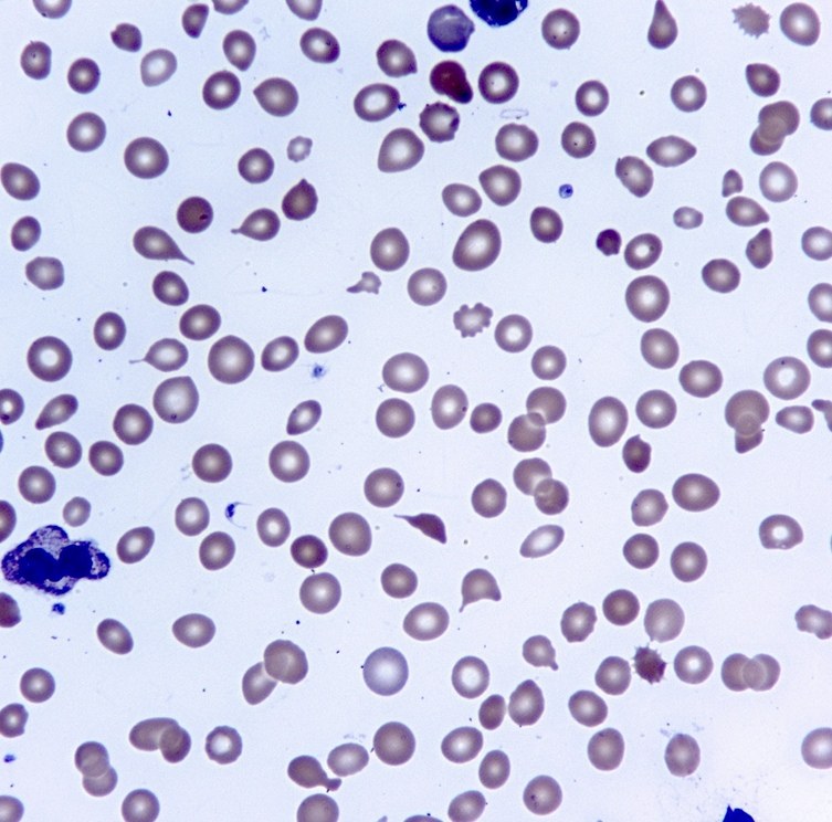
Laboratory diagnostics reveal normochromic normocytic anemia (moderate to severe), accompanied by leucoerythroblastosis with tear-drop cells (dacryocytes) (Fig. 1) and other poikilocytes. The leukocyte count may be significantly elevated in early stages, but is usually normal or decreased in later stages. The platelet count at the time of diagnosis is typically normal or elevated, but thrombocytopenia develops as the disease progresses.
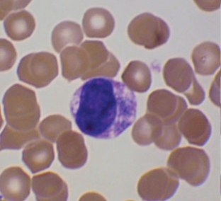
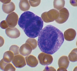
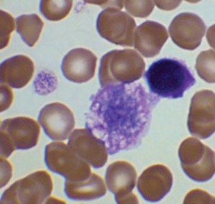
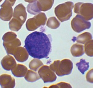
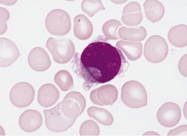
Primary myelofibrosis shows specific changes indicative of its myeloproliferative nature. These include: micromegakaryocytes (Fig. 2), “naked” megakaryocyte nuclei (Fig. 3), giant platelets (Fig. 4), and micromegakaryocytes, budding platelets (Fig. 5). It is often possible to find transitional cells between megakaryoblasts and micromegakaryocytes (Fig. 6). Dysplastic neutrophils also indicate primary myelofibrosis, whereas dyserythropoiesis is much less specific for primary versus secondary myelofibrosis.


