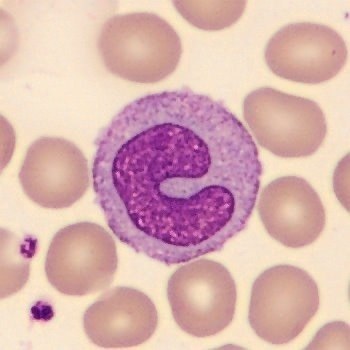
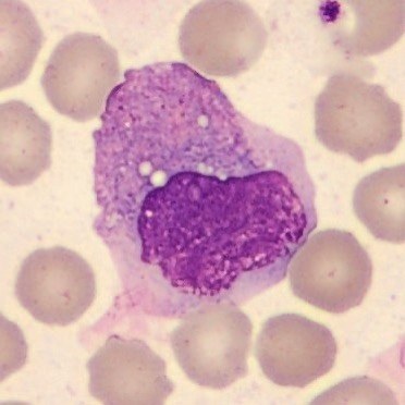
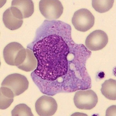
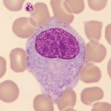
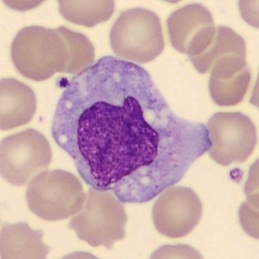
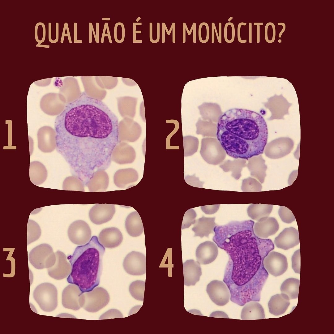
What you NEED to know to correctly identify MONOCYTES? Monocytes are cells with high morphological variability, meaning they are not a cell with a standard shape, but rather an irregular one, similar to reactive lymphocytes (atypical mononuclear cells). To avoid confusing these two cells, pay close attention to two main characteristics: CYTOPLASM and nuclear CHROMATIN. ✅The cytoplasm of a reactive lymphocyte is generally clear, while that of a monocyte is “dusty,” “dirty,” or matte in color. This is because the monocyte cytoplasm has fine granules. Monocyte chromatin is looser and more delicate than that of reactive lymphocytes, which have condensed chromatin with areas of looser chromatin. The nucleus of these cells can sometimes be D-shaped, bean-shaped, or U-shaped, and should not be confused with other cell types that have the same shape, such as metamyelocytes and band neutrophils, respectively. ⚠ Remember: monocytes have gray-blue cytoplasm and looser chromatin. Immature granulocytes have pinkish cytoplasm. ✅Monocytes should also not be confused with blasts, which are very young cells with a high nucleus-to-cytoplasm ratio, loose chromatin, and often a prominent nucleolus. Despite having looser chromatin than other leukocytes, monocyte chromatin is more condensed than that of a blast. 🔑 Pay special attention to chromatin and cytoplasm, which are important clues for confident cell identification 🔐
Monocyte Morphology and Differential Diagnosis in Blood Smears
Understand monocyte morphology: chromatin and cytoplasm assessment, differential diagnosis from reactive lymphocytes and blasts in blood smears.