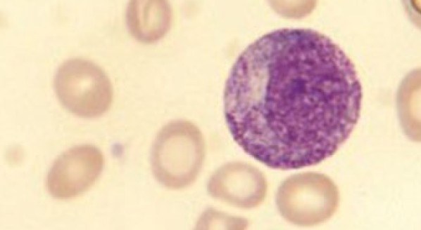
Promyelocyte (Bain B., 2017) Promyelocyte with a lower nucleo-cytoplasmic ratio than a myeloblast, an eccentric nucleus, azurophilic granules, and a Golgi zone to the left of the nucleus. General description from the book. Promyelocytes are rare in the blood of healthy individuals. They are larger than myeloblasts, with more abundant cytoplasm and, consequently, a lower nucleo-cytoplasmic ratio. The cytoplasm is more basophilic than that of a myeloblast and contains azurophilic (pinkish-purple) primary granules. Sometimes, there is a lighter-stained zone in the cytoplasm adjacent to the nucleus, representing the Golgi apparatus, where granules are formed. The nucleus is round or oval, usually eccentric, with some chromatin condensation and a visible nucleolus. Since they lack specific (lineage-associated) granules, promyelocytes, which are precursors to neutrophils, eosinophils, or basophils, usually cannot be distinguished from each other. 💎 Find a large database of photos with descriptions in our Library of Blood and Bone Marrow Secrets. The library volume is over 80 gigabytes! The link with access to the library is automatically sent to you via private message after paying for a VK donut Leukformula subscription.
Promyelocyte Morphology in Peripheral Blood Smear
Morphological features of promyelocytes: nucleo-cytoplasmic ratio, azurophilic granules, Golgi zone, and identification criteria in blood smears.