

Morphological diversity of schistocytes in a 1-year-old boy with hemolytic-uremic syndrome.
Romanowsky-Giemsa stain, ×1000 magnification. 🙏 Material kindly provided by clinical laboratory diagnostics physician Nadezhda Petrenko, Blagoveshchensk.
Morphological features of schistocytes in peripheral blood in a child with hemolytic-uremic syndrome. Romanowsky-stained blood smear microscopy.
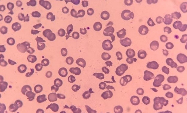
Morphological diversity of schistocytes in a 1-year-old boy with hemolytic-uremic syndrome.
Romanowsky-Giemsa stain, ×1000 magnification. 🙏 Material kindly provided by clinical laboratory diagnostics physician Nadezhda Petrenko, Blagoveshchensk.

Erythrocyte morphology in newborns: detection of Howell-Jolly bodies and binucleated normoblasts in peripheral blood, causes, and evaluation criteria.

A clinical case of mild anemia: severe poikilocytosis, schistocytes, tear-drop cells, and macrothrombocytes in peripheral blood.

Erythrocyte morphology in severe poikilocytosis: detection of ovalocytes, dacryocytes, schistocytes, and stomatocytes in macrocytic anemia and cytopenia.