

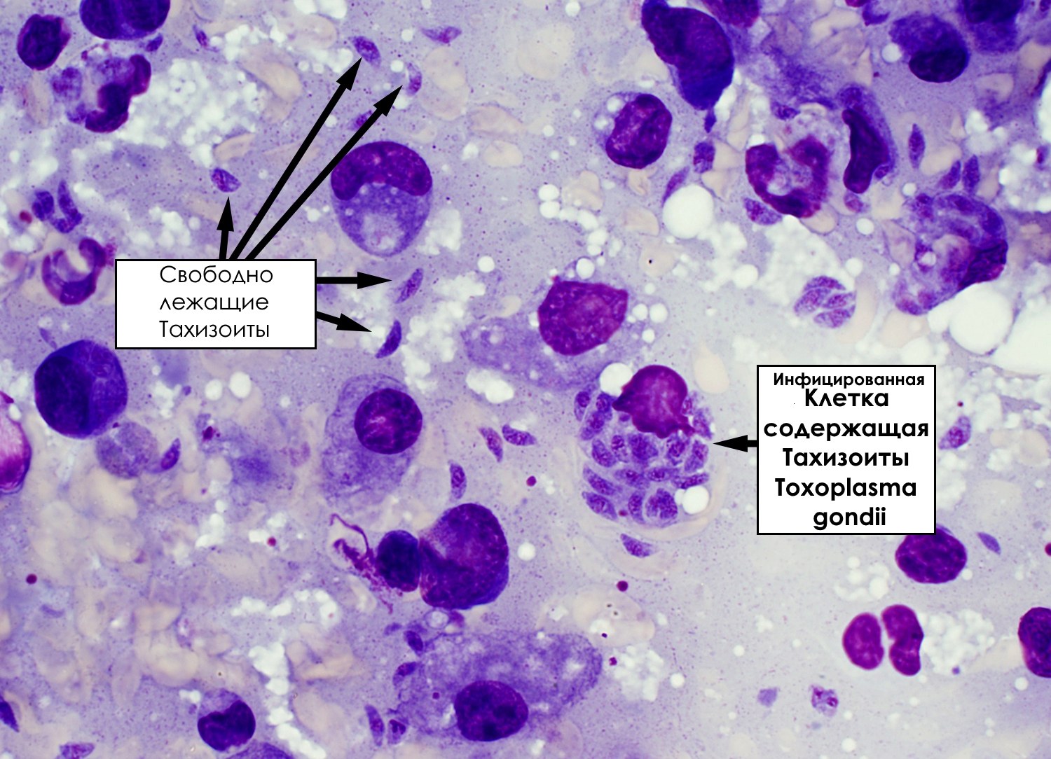
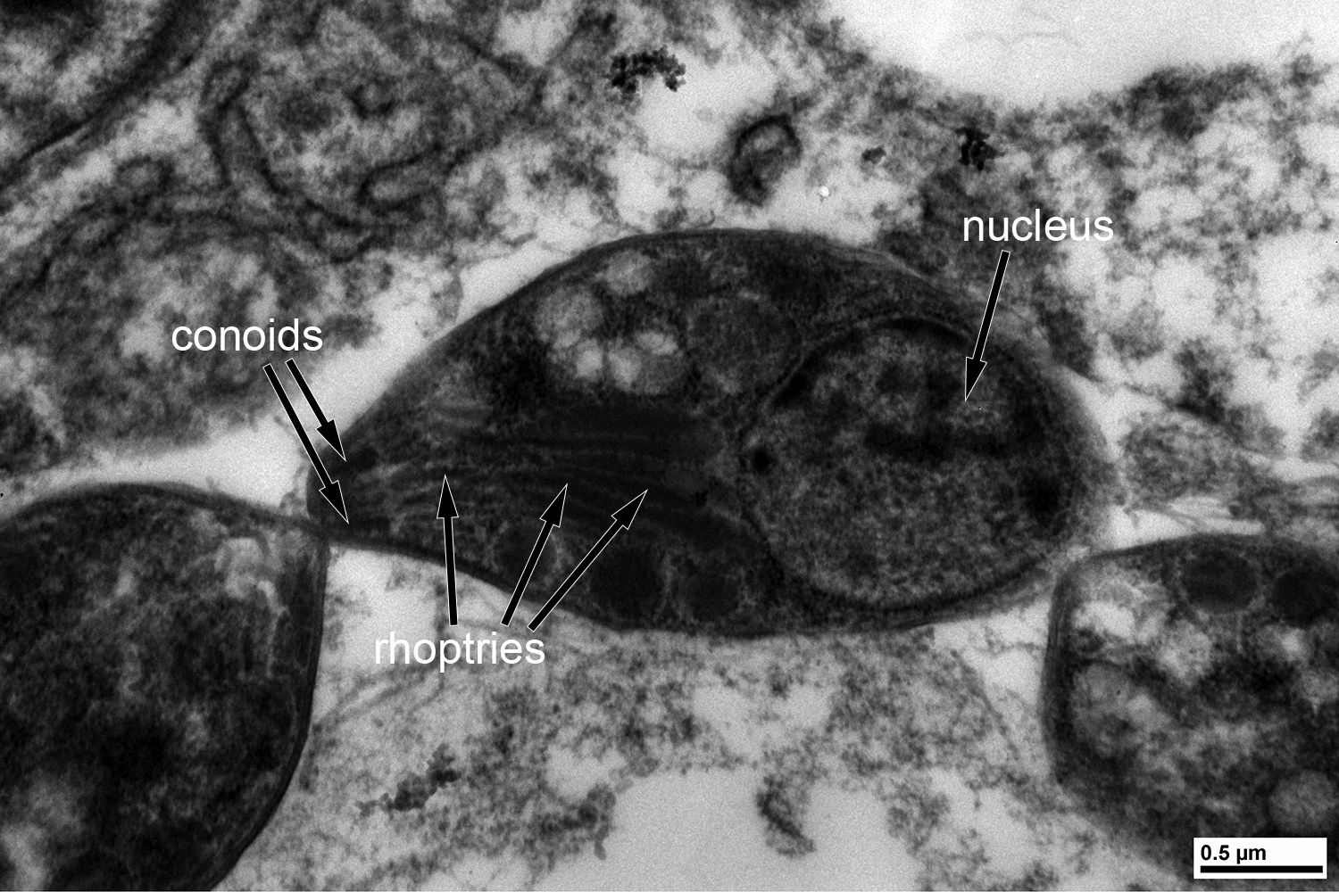
✅ Answer to the parasitology problem: Toxoplasma gondii tachyzoites detected. Among other possible parasites that can be acquired by eating undercooked turkey are Gnathostoma spp., Trichinella pseudospiralis.
Tachyzoites are the rapidly multiplying form of Toxoplasma gondii, characteristic of acute and reactivated infection.
T. gondii can infect any nucleated cell, and tachyzoites are often found both intracellularly and extracellularly during active infection.
Furthermore, the form of tachyzoites visible in smears can vary depending on the tissue fixation method, requiring additional attention for differentiation from other microorganisms such as Histoplasma capsulatum, Trypanosoma cruzi amastigotes, and Leishmania spp.
Now, some fun facts for the curious: Tachyzoites are the rapidly dividing form of T. gondii [tachy comes from Ancient Greek ταχύς (takhús, “fast”)], and are the predominant form observed in acute and reactivated infections. In this case, the presence of numerous extracellular forms is evidence of active infection. In contrast, tissue cysts containing bradyzoites, the slowly replicating forms [brady comes from Ancient Greek βραδύς (bradús, “slow”)], are observed during latent infection. The word zoite comes from Ancient Greek ζῷον (zôion, “animal”). Most medical students know the difference between tachycardia and bradycardia and know that a “zoo” should be thought of as a place where animals live. Helping them apply knowledge they already have helps them learn these new and often very foreign-sounding words.


