

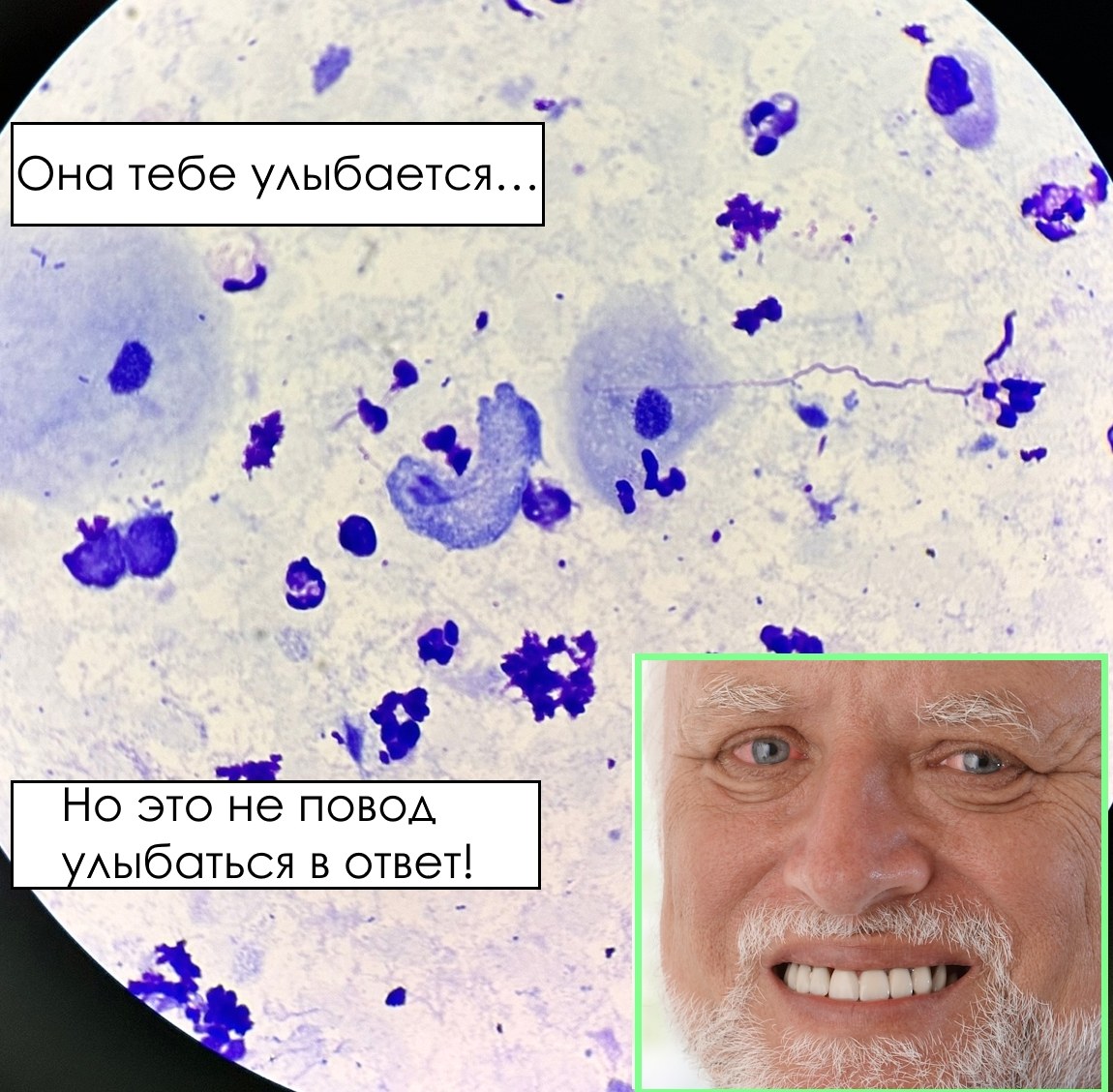
🙏 Image kindly provided by clinical laboratory diagnostics physician, Anna Ivanova, Moscow.
💡 Reference information: Trichomonas (Trichomonas vaginalis) is a single-celled protozoan parasite, the causative agent of trichomoniasis. It inhabits the human urogenital system. It is primarily transmitted through sexual contact. It often has a pear shape and is motile due to flagella. 🔬 Microscopy: In the presented microscopic image of a vaginal smear: the trichomonad appears in a “smile” shape, which is a coincidental visual effect.
✅ Symptoms of trichomoniasis: In women: itching, burning, pathological discharge from the genital tract. In men: less pronounced symptoms (often asymptomatic course).


