

Trichomonas vaginalis in a male flora smear. Confirmed by PCR.
🙏 Images kindly provided by Clinical Laboratory Diagnostics Physician, Irina Selivanova LLC “Citilab-Ural”, Yekaterinburg
Microscopic appearance of Trichomonas vaginalis in a stained male flora smear: leukocyte reaction, epithelial cells, and PCR confirmation.
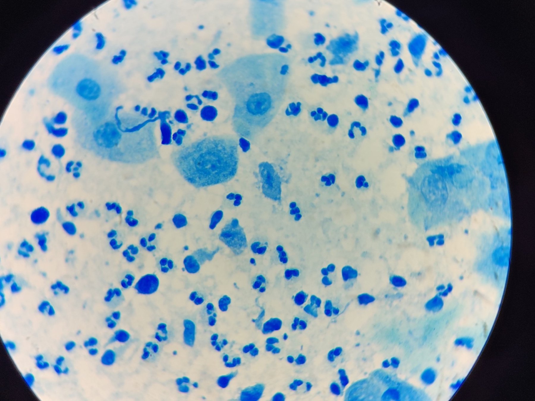
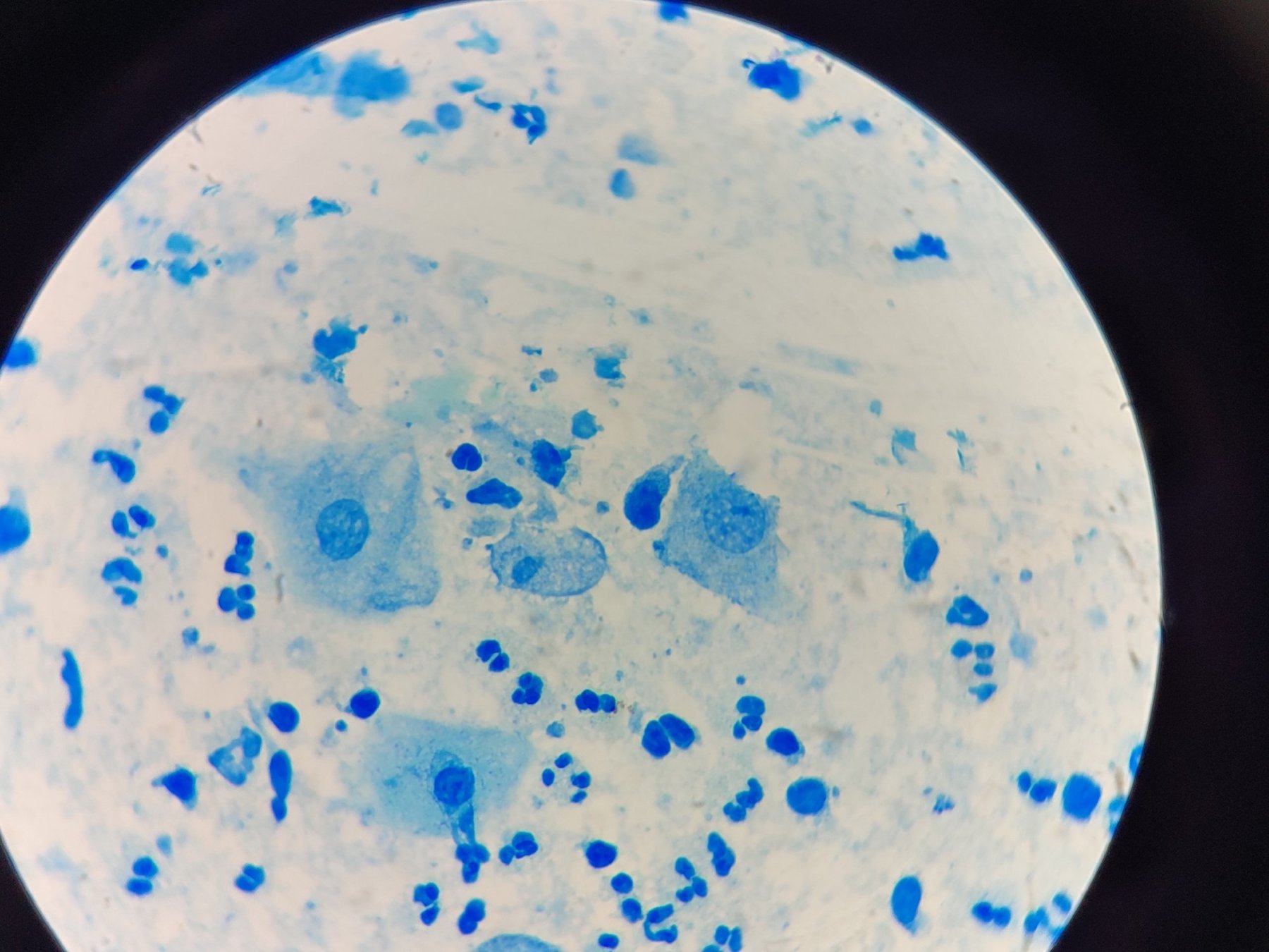
Trichomonas vaginalis in a male flora smear. Confirmed by PCR.
🙏 Images kindly provided by Clinical Laboratory Diagnostics Physician, Irina Selivanova LLC “Citilab-Ural”, Yekaterinburg

Morphology of Trichomonas vaginalis in stained urogenital smears: identification of protozoa, leukocyte reaction, and bacterial flora during microscopy.

Microscopy of a stained gynecological smear: detection of Trichomonas vaginalis with flagella amidst abundant bacterial flora and leukocytes.

Learn the morphology of Trichomonas vaginalis during microscopy of stained urogenital smears, including criteria for detection and the cytological picture.