

Trichomonads in a female smear. Eccentrically located nuclei, axostyle, and in some, anterior and posterior flagella are visible.
🙏 The publication was kindly provided by KDL physician Nana Novikova, CHUZ “KB” RZHD-Meditsina" Smolensk.
Photos
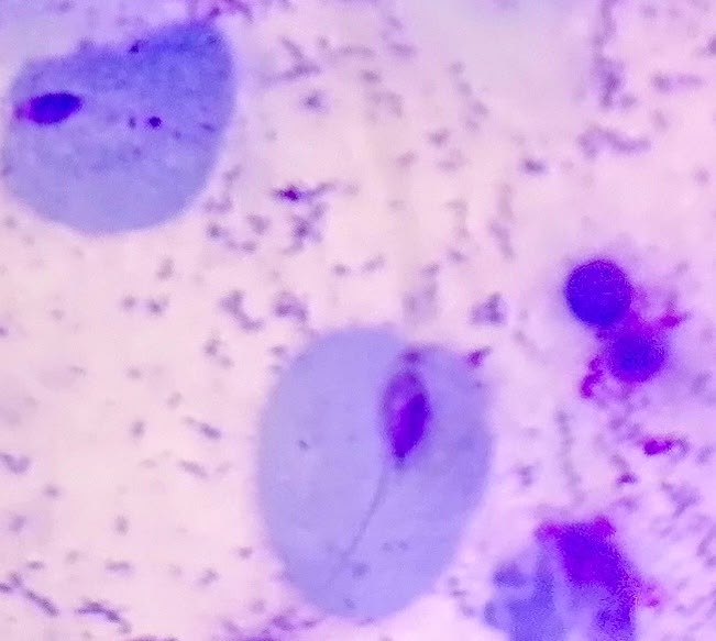
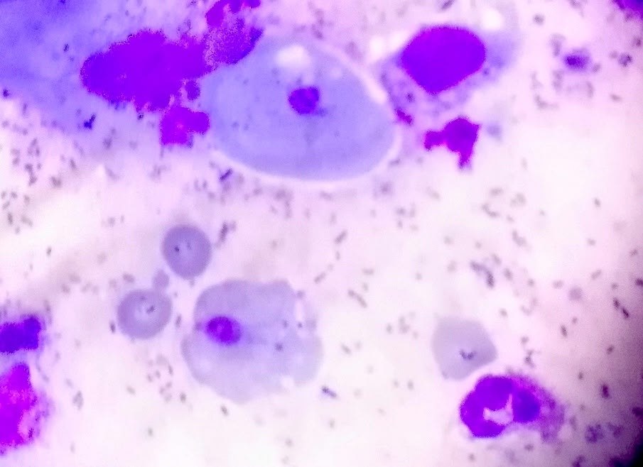
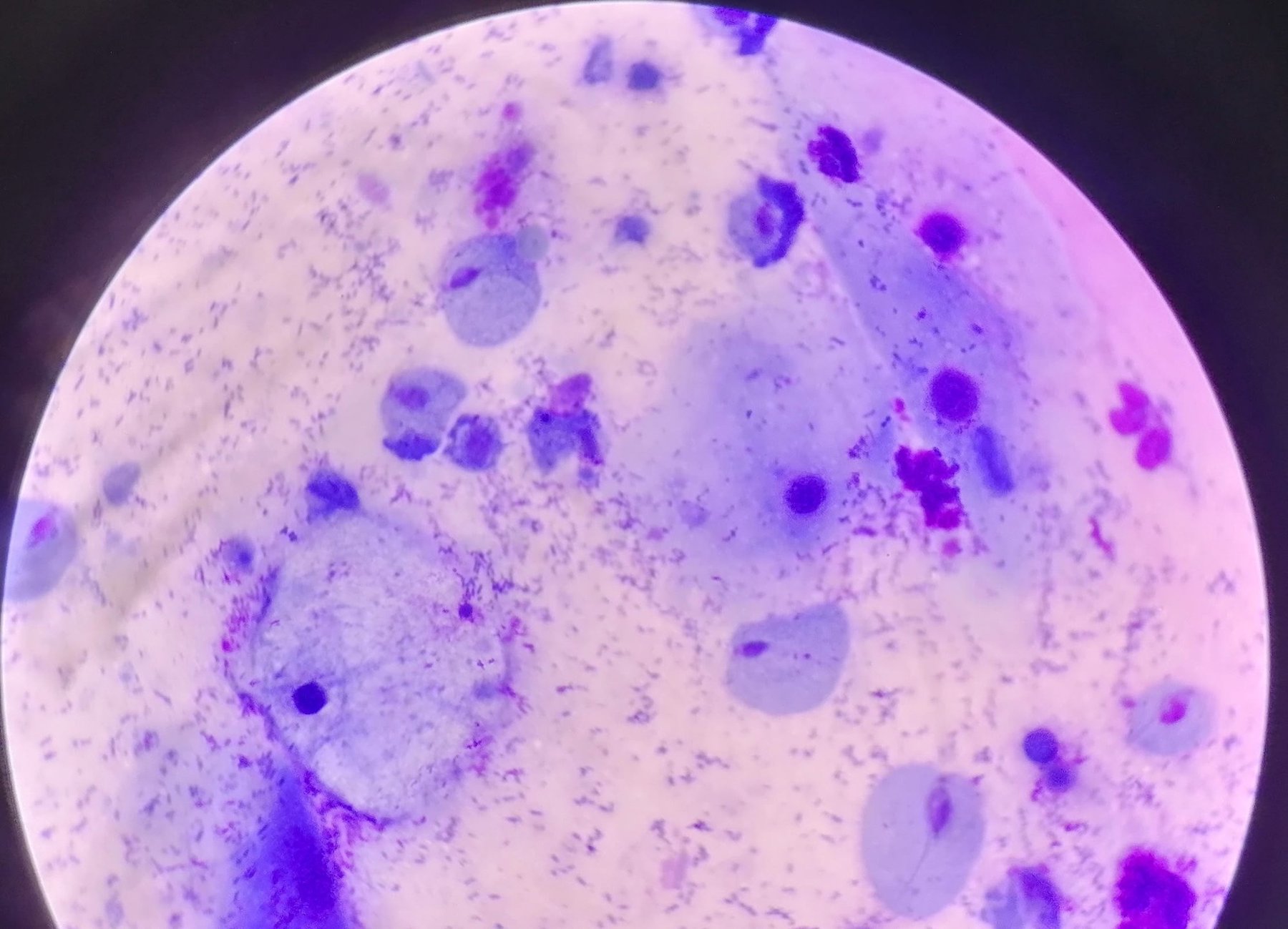
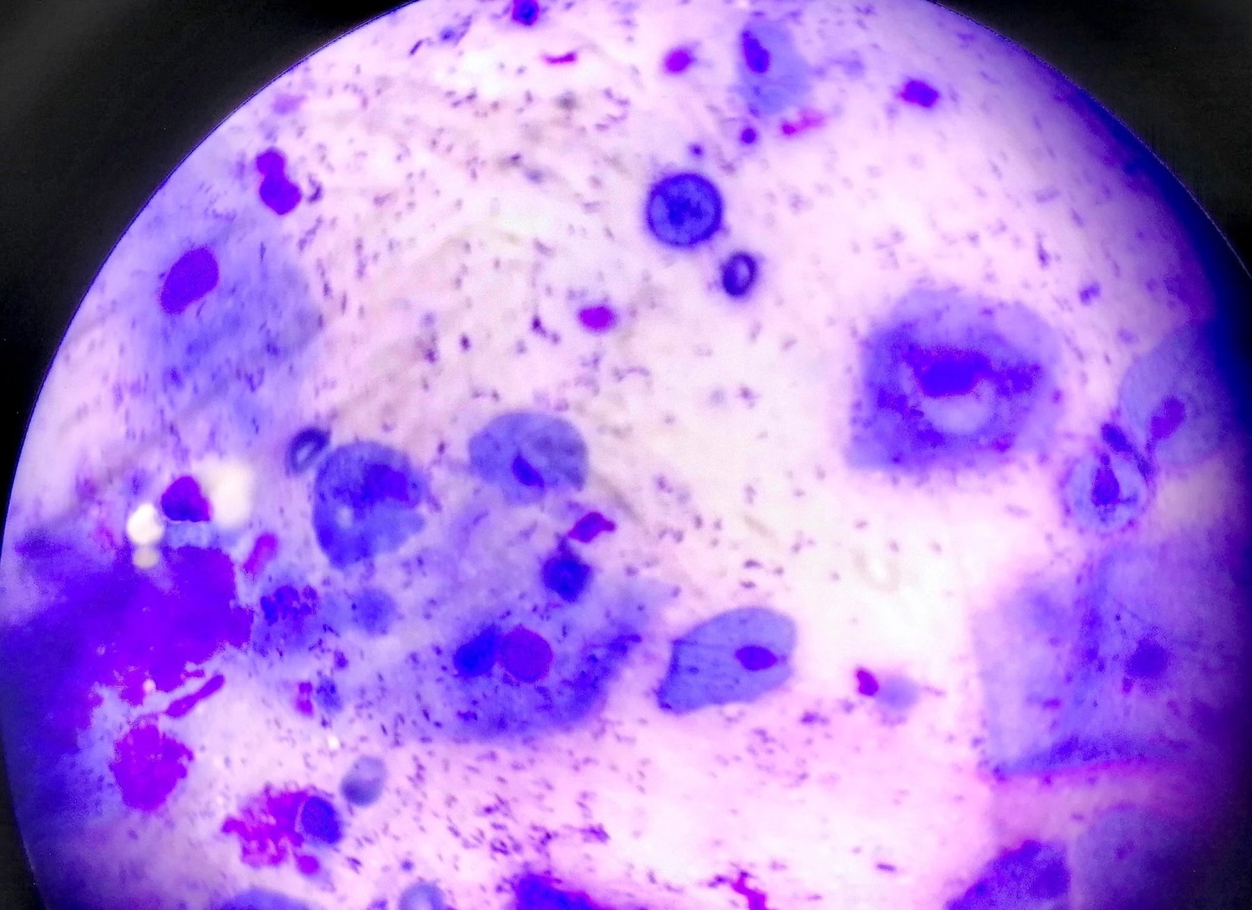
Microscopic appearance of Trichomonas vaginalis in a stained smear: eccentric nuclei, axostyle, and flagella of protozoa amidst epithelium and microflora.
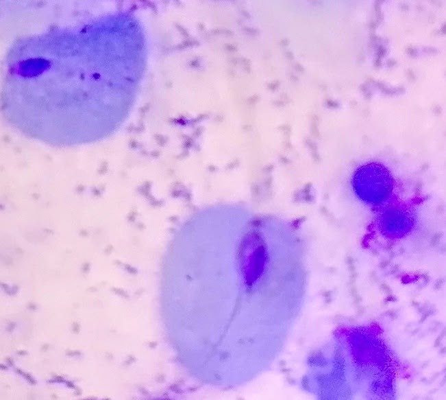
Trichomonads in a female smear. Eccentrically located nuclei, axostyle, and in some, anterior and posterior flagella are visible.
🙏 The publication was kindly provided by KDL physician Nana Novikova, CHUZ “KB” RZHD-Meditsina" Smolensk.

Morphology of Trichomonas vaginalis in stained urogenital smears: identification of protozoa, leukocyte reaction, and bacterial flora during microscopy.

Microscopic view of a urogenital smear: identifying Trichomonas vaginalis trophozoites among squamous epithelial cells and abundant microflora.

Microscopic appearance of Trichomonas vaginalis in a stained male flora smear: leukocyte reaction, epithelial cells, and PCR confirmation.