
🔬 Trichomonas, urogenital smear. Stained with Romanowsky-Giemsa.
🙏 Photos kindly provided by laboratory physician Viktoriya Gerbutova, Krasnodar.
Microscopic appearance of Trichomonas vaginalis in a urogenital smear stained with Romanowsky-Giemsa: flagella, nucleus, and characteristic cytoplasm.
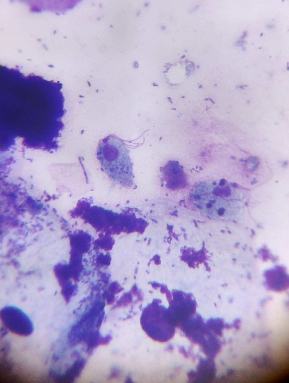
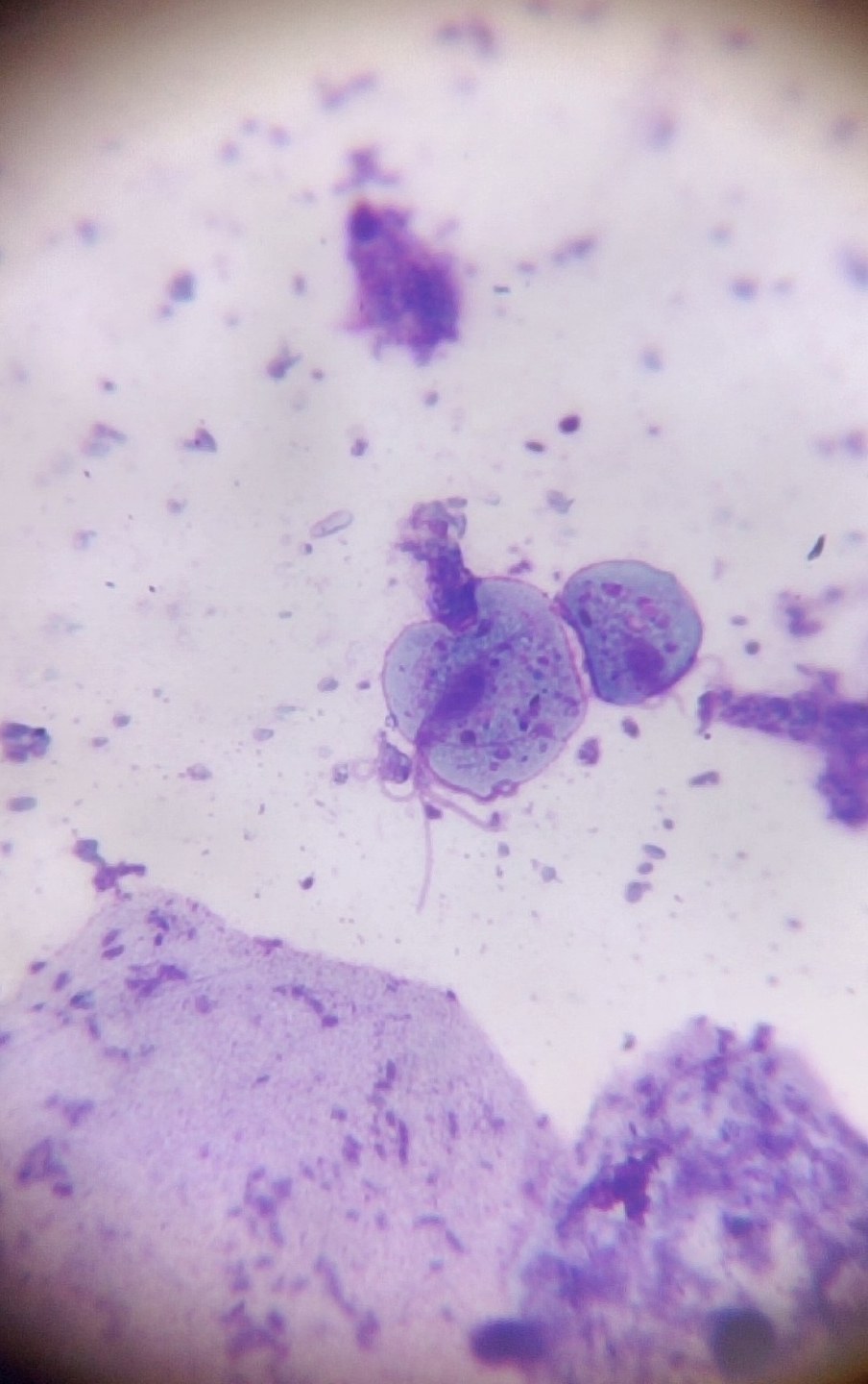
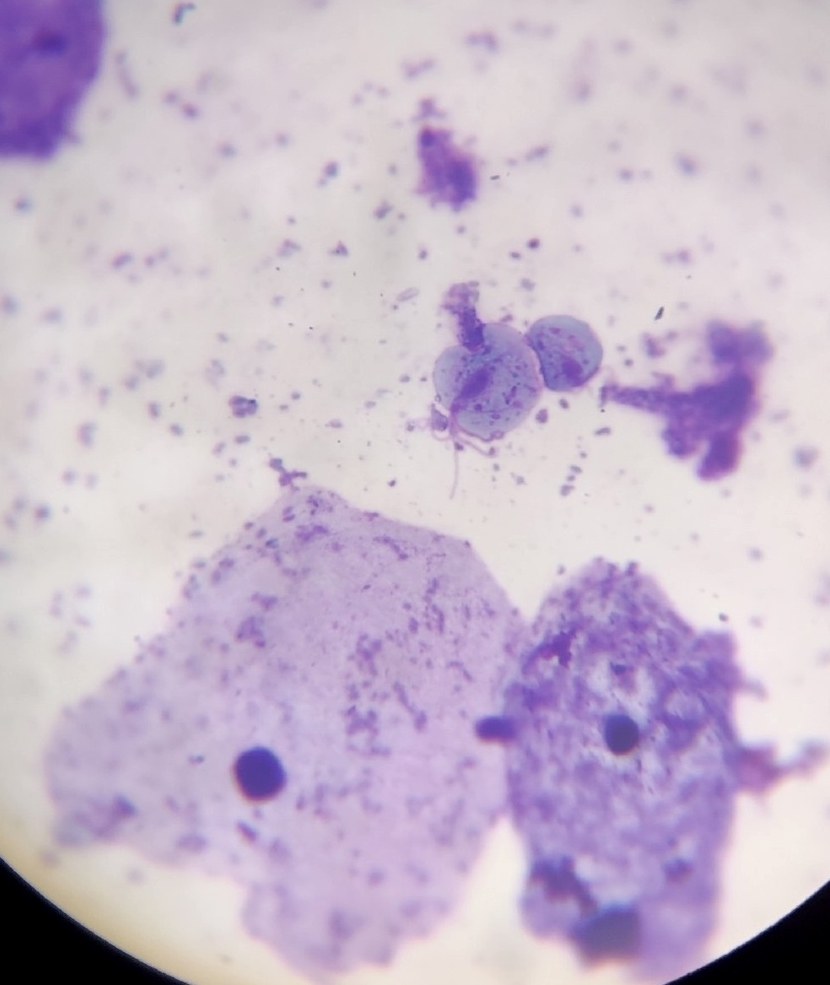
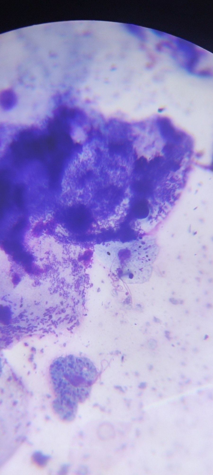
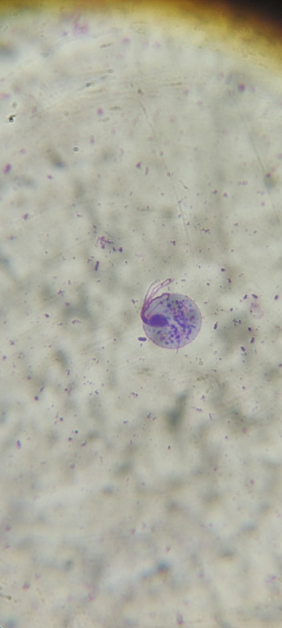
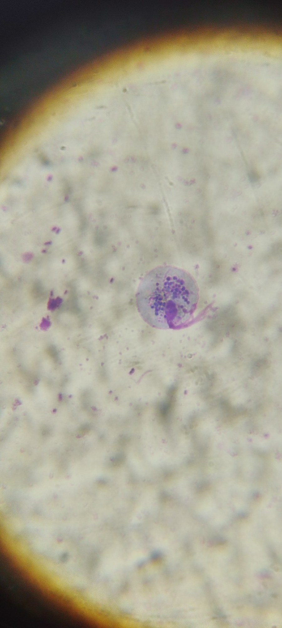
🔬 Trichomonas, urogenital smear. Stained with Romanowsky-Giemsa.
🙏 Photos kindly provided by laboratory physician Viktoriya Gerbutova, Krasnodar.

Morphological features of Trichomonas vaginalis in a flora smear stained with Romanowsky stain: detection of trophozoites amidst leukocyte infiltration.

Microscopic features of urogenital trichomoniasis. Specific morphological characteristics of Trichomonas vaginalis trophozoites stained with Romanowsky-Giemsa.

Microscopy of Trichomonas vaginalis in a gynecological smear stained by Romanowsky method: detection of trichomonad trophozoites, leukocyte reaction, and flora.