
❓How many trichomonads can you count?
🙏 The image was kindly provided by Clinical Laboratory Diagnostics physician Victoria Gerbutova, Krasnodar.
Microscopic view of a urogenital smear: detection of Trichomonas vaginalis, morphological features of protozoa, and differential diagnosis.
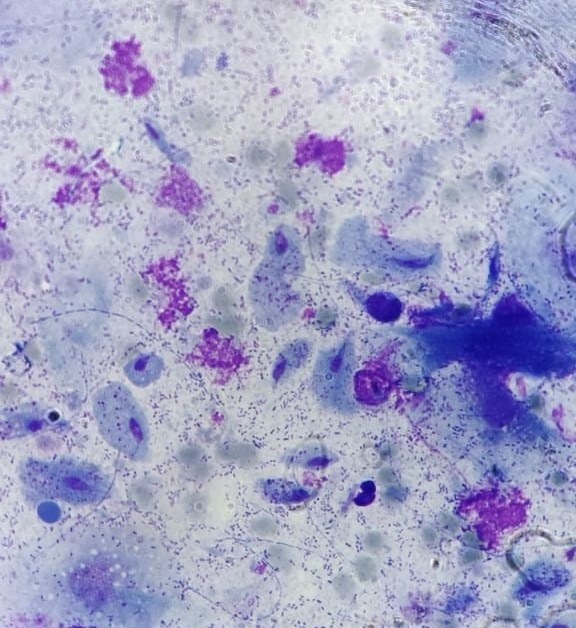
❓How many trichomonads can you count?
🙏 The image was kindly provided by Clinical Laboratory Diagnostics physician Victoria Gerbutova, Krasnodar.

Microscopic appearance of Trichomonas vaginalis in a urogenital smear stained with Romanowsky-Giemsa: flagella, nucleus, and characteristic cytoplasm.

Microscopic view of a urogenital smear: identifying Trichomonas vaginalis trophozoites among squamous epithelial cells and abundant microflora.

Microscopic appearance of Trichomonas vaginalis in a stained male flora smear: leukocyte reaction, epithelial cells, and PCR confirmation.