

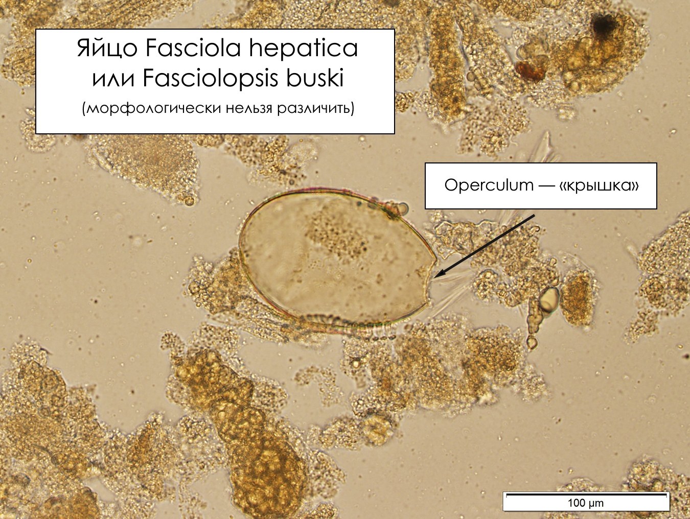
🐍 Correct answer to the parasitology problem. 👉 Fasciola hepatica or Fasciolopsis buski eggs. ✅ The conclusion can be made based on the large size of the egg (>100 micrometers in diameter) and the presence of an operculum. Size is an important feature for differentiating F. hepatica/F. buski eggs from Diphyllobothrium and Paragonimus eggs, which are similar in appearance but smaller. Unfortunately, it is not possible to reliably differentiate F. hepatica and F. buski eggs based on morphology alone.
✅ The image below from this case clearly shows where the operculum is located. It is often difficult to see the operculum on Fasciola/Fasciolopsis eggs because it lacks shoulders and often blends with the egg wall. One way to see the operculum is to gently press on the coverslip and thereby open it! 💎 Look for more useful materials on parasitology in our LAB-TESTS library. The link to access the library is automatically sent to you via private message after paying for a VK donut Labformula subscription.


