

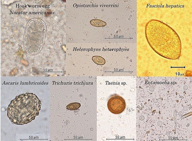
Collection of parasite eggs.
Necator americanus — Egg of an intestinal helminth (roundworm) causing hookworm infection. Characteristically oval egg.
Opisthorchis viverrini — Egg of a liver fluke causing opisthorchiasis. Small, oval, with a characteristic “tail” at one end.
Heterophyes heterophyes — Egg of another type of fluke, parasitic in the intestine. Similar to Opisthorchis.
Fasciola hepatica — Egg of the liver fluke. Larger in size, oval, with a distinctly prominent operculum at one end.
Ascaris lumbricoides — Egg of Ascaris, a large roundworm causing ascariasis. Characterized by a rough surface and a thick shell.
Trichuris trichiura — Small in size.
Taenia sp. — Egg of beef or pork tapeworm (e.g., Taenia solium or Taenia saginata).
Entamoeba sp. — Protozoan parasite


